INTRODUCTION
Even today,
tuberculosis (TB) represents a significant global public health problem, being
the tenth leading cause of death worldwide and the leading cause of death from
a single infectious agent. Deaths in the pediatric population account for
approximately 15% of the total.1
Mortality rates
from untreated TB can reach 14.9% in children aged 5 to 14 years, and 43.6% in
children under 5 years of age. Despite its severity, many cases of childhood TB
are underreported due to diagnostic difficulties. With timely diagnosis and
treatment, mortality can be reduced to as low as 0.9%.2,3 Although
pulmonary disease is the most common form of presentation, the pediatric
population may present with extrapulmonary or disseminated disease in about
20-30% of cases.4
Central nervous
system tuberculosis (CNS TB) is a less frequent, but serious, form of
extrapulmonary involvement, with high mortality rates and neurological
sequelae. It occurs in up to 5 to 10% of cases, with children under 5 years of
age at higher risk.4-6 Its often nonspecific symptomatology
contributes to diagnostic and therapeutic delays, leading to a high risk of
death and other unfavorable outcomes.6
Thus, raising
awareness among healthcare professionals to recognize possible signs and
symptoms of CNS TB is essential to improve the prognosis of patients affected
by the disease. Therefore, this article aims to describe a case of CNS TB in a
pediatric patient, documenting clinical manifestations, complementary
examinations, diagnosis, management, and main outcomes.
CASE DESCRIPTION
This is a
report of a 10-year-old male patient, initially referred from a hospital in the
interior of the state for evaluation by the institution’s oncology service in
September 2022, due to suspected posterior fossa (PF) brain tumor. In his
personal medical history, he had an up-to-date vaccination schedule for his age
group according to the National Immunization Program and no associated
comorbidities.
During the two
months preceding the referral, the patient sought medical attention several
times for daily, intense headaches in the right parieto-occipital region,
associated with general malaise, prostration, drowsiness, blurred vision, loss
of appetite, and vomiting preceded by nausea. He did not respond to simple
analgesia. He also presented with daily afternoon and night fever, associated
with intense sweating. Eventually, he was referred to a tertiary pediatric
hospital in the interior of the state due to signs of intracranial hypertension
(ICH) observed on cranial computed tomography (CT) scan. The image showed
dilation of the ventricular system associated with hypoattenuation of the
periventricular cerebral parenchyma, suggesting hydrocephalus with hypertensive
signs, and apparent effacement of cerebral sulci, suggesting edema. For this
reason, during hospitalization, surgery was performed to insert an external
ventricular drain (EVD), later transformed into a ventriculoperitoneal shunt
(VPS). The patient remained hospitalized in the Intensive Care Unit (ICU) for 5
days after the procedure.
The possibility
of a PF brain tumor was raised by the originating hospital, and the patient was
referred to the oncology service at the tertiary hospital in Florianópolis. In
the week preceding the transfer, the patient also presented with a tonic-clonic
seizure, lasting approximately five minutes, and began with transient episodes
of left hemiparesthesia and asymmetry of facial expression.
At the time of
admission to the referral center, the patient presented on physical examination
in good general condition and with a good overall impression, communicative,
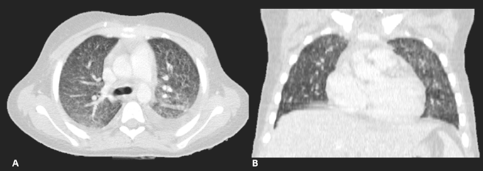
slightly pale (+/4) and slightly tachypneic. Cardiac auscultation was
unremarkable; pulmonary auscultation revealed reduced vesicular murmurs on the
left with fine crackles at the base. Abdomen diffusely painful on deep
palpation without other abnormalities. Extremities well perfused, pulses strong
and symmetrical, and capillary refill time of 3 seconds, lower limbs (LL) with
cold and symmetrical edema up to the ankles, calves free. On neurological
examination, facial expression was asymmetrical and strength was symmetrically
reduced in the LL, without gait impairment.
The patient was
admitted for investigation, this being day 1 (D1) of hospital admission.
Laboratory tests were requested, which revealed microcytic and hypochromic
anemia (hemoglobin 10.1 g/dL; MCV 72 fL; MCH 25 pg); elevated inflammatory
markers (CRP 8.9 mg/L; ESR 20 mm/h); hyponatremia and hypokalemia (Na 129
mEq/L; K 3.0 mEq/L); alterations in the coagulation profile (PT 17.09 seconds;
INR 1.36; aPTT 39 seconds); slightly altered liver function (AST 66 IU/L; ALT
43 IU/L). The other parameters were unchanged.
In addition to laboratory
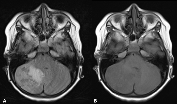
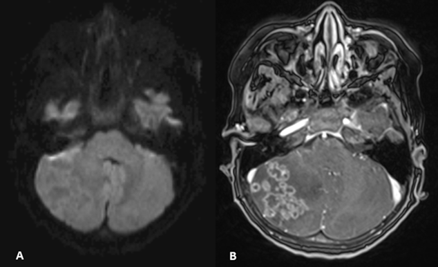
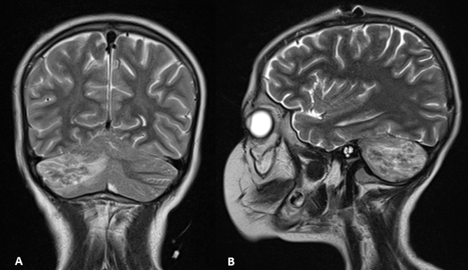
follow-up, on D5, a neuroaxial magnetic resonance imaging (MRI) scan was
performed (Figures 1, 2, and 3), which showed multiple cystic lesions with ring
enhancement, intra-axial and supra- and infratentorial leptomeningeal, in
greater number and of larger dimensions in the right cerebellar hemisphere,
causing vasogenic edema in the adjacent brain parenchyma, with consequent local
mass effect, in addition to occipital and frontal (subcortical) lesions of a
similar pattern and without diffusion restriction. No spinal cord involvement
was evident. Based on the findings, the hypotheses of leptomeningeal neuroglial
tumor or inflammatory/infectious disease, mainly granulomatous, were raised.
On D8, cerebrospinal fluid
(CSF) was collected for analysis, with the following results: nucleated cells
85/mm3 with a lymphocytic predominance of 62%; red blood cells 5/mm3; glucose
31 mg/dL; protein 120 mg/dL; negative acid-fast bacilli (AFB); detection of
traces of Mycobacterium tuberculosis DNA through a rapid molecular test
(GeneXpert® MTB/RIF Ultra); negative cultures for fungi and Cryptococcus.
Pleural fluid was also evaluated, with the following results: glucose 85 mg/dL;
nucleated cells 230/mm3 (lymphocytes 67%; neutrophils 30%; basophils 1%);
protein 2.0 g/dL (albumin 1.0 g/dL; globulin 1.0 g/dL); red blood cells 65/mm3;
negative AFB. TRM for Mycobacterium tuberculosis was not detected in
pleural fluid or pleural biopsies. The HIV serology result was non-reactive.
Thus,
disseminated TB with CNS involvement was diagnosed, and on D10, a standardized
regimen recommended by the Ministry of Health (MH) was initiated, with RHZE
(rifampicin, isoniazid, pyrazinamide, and ethambutol) to be maintained for 2
months, followed by 10 months of RH (rifampicin and isoniazid), totaling 12
months of treatment. Intravenous corticosteroids were also initiated as per MH
guidelines for severe CNS TB, later transitioned to oral therapy.
After the
introduction of medication, the patient showed progressive improvement in
symptoms, remaining afebrile. There was improvement in neurological changes,
with only blurred vision remaining, in addition to improvement in laboratory
parameters, with only anemia remaining.
As
complications, he presented with tonic-clonic seizures, requiring management
with anticonvulsant medication (phenytoin), and thrombosis of the right
external iliac vein, managed with warfarin. He was discharged from the hospital
16 days after starting treatment with RHZE. At the time of discharge, he was
using phenytoin 100mg every 12 hours, warfarin 8.5mg/day, in addition to RHZE,
and received referrals to hematology, neurology, and neurosurgery services for
outpatient follow-up.
The patient
used warfarin for 9 months and phenytoin for 8 months, with subsequent
discontinuation without recurrence of thromboembolic events or seizures, and
was discharged from the hematology and neurology outpatient clinics. In the
latest neurological assessment, the patient presented preserved visual field
and oculomotor function, without nystagmus, normal facial expression, no
dysmetria, and no meningeal signs. Strength was preserved in the lower limbs
and upper limbs, with an atypical gait. However, he presents inattentive and
anxious behavior, sometimes hetero-aggressive, and has started follow-up with a
psychiatrist and psychologist.
The patient was
discharged from the infectious disease outpatient clinic after 12 months of
treatment with excellent adherence to RHZE, showing good tolerance to the
medication. He showed complete improvement of symptoms, remaining without
neurological or pulmonary alterations.
DISCUSSION
CNS TB
represents a severe form of extrapulmonary TB, with high mortality rates and
neurological sequelae, such as motor deficits and cognitive disorders. It shows
pleomorphic clinical pictures, and early diagnosis is directly related to the
prevention of negative outcomes by providing timely treatment. Thus, high
clinical suspicion is fundamental, especially in the pediatric population, in
which 5 to 10% of TB cases may present with CNS involvement.7
Among the main
symptoms of CNS TB are headache, vomiting, and altered mental status.
Hydrocephalus, intracranial hypertension, and seizures may also be found,6
in addition to symptoms compatible with cranial nerve involvement – mainly
oculomotor, optic, and abducens nerves – such as paralysis and focal deficits
related to the functionality and topography of each nerve.8 In the
case described, all these clinical findings were present, in addition to
afternoon fever and night sweats, symptoms highly suggestive of TB. Given this
clinical picture, it is imperative that the diagnosis of TB be considered,
especially given its high prevalence and importance to public health in Brazil.5
In the case described, a delay in clinical suspicion was observed, with
oncological diagnoses considered rare initially being considered. Diagnostic
delay in similar cases represents a failure that can compromise the prognosis.9
Regarding
laboratory investigation, cerebrospinal fluid analysis stands out, which, when
suggestive of CNS TB, shows pleocytosis with lymphocytic predominance, a slight
increase in protein levels, and low glucose levels, a pattern compatible with
that presented in the clinical case. In addition, because it contains few
bacilli, it is expected that it will be difficult to observe M. tuberculosis
in the CSF by direct method, and molecular techniques should be used to detect
genetic material, which have good accuracy for detecting the bacillus in the
CSF. In the reported case, the nucleic acid amplification technique was
performed with the GeneXpert® MTB/RIF Ultra test. Despite the still nascent
literature, studies indicate that this molecular methodology performs
adequately for the diagnosis of extrapulmonary TB in paucibacillary tissues –
such as CNS TB – and provides information on rifampicin resistance,
contributing to better therapeutic management.10
In addition to
cerebrospinal fluid alterations, there may be other laboratory changes, the
most frequent being: anemia, leukocytosis, elevation of inflammatory markers
and hydroelectrolytic disorders, with emphasis on hyponatremia – usually related
to aggravating conditions such as salt-wasting brain syndrome and syndrome of
inappropriate antidiuretic hormone secretion – and hypocalcemia.11,12
These findings are not very sensitive or specific for TB and CNS TB.13
With respect to
the radiological findings of CNS TB, the most common are hydrocephalus, basal
meningeal thickening and cerebral infarcts. Parenchymal involvement, manifested
by the presence of tuberculomas and brain abscesses, usually results from
hematogenous dissemination, justifying the fact that they are frequently found
in association with other sites of TB infection.6 The radiological
characteristics of tuberculomas often mimic those of other infectious and
non-infectious conditions, such as neurocysticercosis, metastasis, CNS lymphoma,
neurotoxoplasmosis, tumors, and pyogenic abscess. Therefore, other diagnostic
methods besides CT – such as MRI – should be used for better diagnostic
clarification.14
In cases of
parenchymal involvement, tuberculomas are the most frequent lesions, and occur
due to the coalescence of microgranulomas.13 In children, the
infratentorial regions are the most affected, especially the PF.6 In
non-contrast CTs, tuberculomas may present as iso- or hypodense masses, while
contrast-enhanced CTs and MRIs demonstrate solid lesions with ring enhancement.
The most frequently described neuroimaging findings are like those found in the
reported case.13 The patient also presented signs of
non-communicating hydrocephalus, a relatively frequent form of presentation of
CNS TB that results from obstructions and mass effect caused by the presence of
the tuberculoma.13
Because it is a
severe form of extrapulmonary involvement, which is associated with high
morbidity and mortality and a risk of permanent neurological sequelae, it is
imperative that, upon suspicion of a diagnosis of CNS TB, treatment be promptly
initiated.6,15 The Ministry of Health recommends treatment for 12
months, using the RHZE regimen for the first 2 months, followed by 10 months of
the RH regimen (2RHZE/10RH regimen).15 In addition, the use of
systemic corticosteroid therapy is strongly recommended, as it has been shown
to reduce morbidity and mortality rates and disease recurrence, with the
benefit being greater the higher the severity of the case. The importance of
adequate treatment with RHZE at the time of implementation of corticosteroid
therapy is emphasized, to mitigate the risks of immunosuppression and worsening
of the condition.15
The prognosis
for CNS TB cases tends to be unfavorable, with high mortality rates and
permanent neurological sequelae. In children, the main factors associated with
a worse prognosis are young age, lower socioeconomic levels, presence of
altered mental status and level of consciousness, abnormal postures,
hydrocephalus, and basal exudates.8 However, with appropriate
treatment, it is possible to progress to clinical improvement and complete
remission of neurological symptoms, as observed in this patient.
Therefore, the
importance of surveillance for CNS TB in children is reinforced, especially in
the presence of warning signs and in regions with a high prevalence of TB, such
as Brazil. Suspicion of CNS TB should be considered in children with
neurological symptoms of subacute and protracted evolution, especially when
accompanied by systemic manifestations and nonspecific findings in
complementary examinations. As observed in this case, differential diagnosis is
fundamental, although it may result in delays in diagnostic definition and
untimely interventions. Raising awareness among health professionals is
essential to reduce delays in diagnosis, enabling the implementation of early
treatment and ensuring a better prognosis for patients affected by the disease.
REFERENCES
1.
World Health Organization (WHO). WHO consolidated guidelines on
tuberculosis (Module 1 – Prevention): Tuberculosis preventive treatment, second
edition. Geneva, World Health Organization. 2024. Avaliable from:
https://www.who.int/publications/i/item/9789240096196. Accessed March 26, 2025.
2.
Jenkins HE, Yuen CM, Rodriguez CA, Nathavitharana RR, McLaughlin
MM, Donald P, et al. Mortality in children diagnosed with tuberculosis: a
systematic review and meta-analysis. Lancet Infect Dis [Internet]. 2017
Mar;17(3):285-95. Avaliable from: https://www.sciencedirect.com/science/article/pii/S1473309916304741.
Accessed
March 26, 2025.
3.
World Health Organization (WHO). WHO consolidated guidelines on
tuberculosis (Module 5 - Management of tuberculosis in children and
adolescents). Geneva, World Health Organization. 2022. Avaliable from:
https://www.who.int/publications/i/item/9789240046764. Accessed March 26, 2025.
4.
Maphalle LNF, Michniak-Kohn BB, Ogunrombi MO, Adeleke OA.
Pediatric Tuberculosis Management: A Global Challenge or Breakthrough? Children
[Internet]. 2022 Jul 27;9(8):1120. Avaliable from:
https://www.ncbi.nlm.nih.gov/pmc/articles/PMC9406656/. Accessed March 26, 2025.
5. Cortez AO, Melo AC, Neves
LO, Resende KA, Camargos P. Tuberculosis in Brazil: one country, multiple
realities. J Bras Pneumol [Internet]. 2021 Apr 30;47(2):e20200119. Avaliable from:
https://www.scielo.br/j/jbpneu/a/DsDmc6KJFtcCxG8tfkBcGLz/?lang=pt&format=pdf.
Accessed May 15, 2025.
6.
Jamoussi M, Benrhouma H, Miladi Z, Ben Younes T, Klaa H, Rouissi
A, Kraoua I, Ben Youssef I. Pediatric Neurotuberculosis: A cases series and
review of the literature. Eur J Paediatr Neurol [Internet]. 2023 Mar; 43: 6-11.
Avaliable from: https://pubmed.ncbi.nlm.nih.gov/36739787/. Accessed March 26, 2025.
7. Ogeda P, Tedesco R,
Cavalli C, Piasecki V, Piasecki T, Berto L, et al. Neurotuberculosis in
childhood: a case report. Residência Pediátrica [Internet]. 2024;14(3).
Avaliable from:
https://cdn.publisher.gn1.link/residenciapediatrica.com.br/pdf/v14n3e1135.pdf.
Accessed May 15, 2025.
8. Wen A, Cao WF,
Liu SM, Zhou YL, Xiang ZB, Hu F, et al. Incidence and Risk Factors of Cranial
Nerve Palsy in Patients with Tuberculous Meningitis: A Retrospective
Evaluation. Infection and Drug Resistance [Internet]. 2023 Feb 1; 16:829-41.
Avaliable from: https://pmc.ncbi.nlm.nih.gov/articles/PMC9938703/
9.
Sharma V, Rajeshwari K, Kumar D, Gupta G. Clinicoepidemiological
Profile and Prognostic Factors in Neurotuberculosis in Children. Ann Child
Neurol. 2023 Apr 1;31(2):103-12. Avaliable from:
https://www.annchildneurol.org/journal/view.php?number=1314. Accessed May 15, 2025.
10. Kay AW, Ness T, Verkuijl
SE, Viney K, Brands A, Masini T, et al. Xpert MTB/RIF Ultra assay for
tuberculosis disease and rifampicin resistance in children. Cochrane Database
of Systematic Reviews. 2022 Sep 6;2022(9). Avaliable from: https://www.cochranelibrary.com/cdsr/doi/10.1002/14651858.CD013359.pub3/full
11.
Ufoaroh CU, Onwurah CA, Mbanuzuru VA, Mmaju CI, Chukwurah SN,
Umenzekwe CC, et al. Biochemical changes in tuberculosis. PAMJ [Internet].
2021;38. Avaliable from: https://pubmed.ncbi.nlm.nih.gov/33889232/. Accessed May 15,
2025..
12.
Jafari NJ, Izadi M, Farhad S, Heidari A, Ranjbar R, Amin S.
Hyponatremia due to pulmonary tuberculosis: Review of 200 Cases. Nephro-Urology
Monthly [Internet]. 2012 Dec 15. Avaliable from: https://pmc-ncbi-nlm-nih-gov.translate.goog/articles/PMC3614323/?_x_tr_sl=en&_x_tr_tl=pt&_x_tr_hl=pt&_x_tr_pto=tc. Accessed September 29,
2025.
13. Agarwal S, Bheru DC, Ahmad
S, Singh E, Garg A. Imaging in Neurotuberculosis. Indographics [Internet]. 2024
Aug 14;03(02):135-49. Avaliable from:
https://www.thieme-connect.com/products/ejournals/pdf/10.1055/s-0044-1788998.pdf.
Accessed May 15, 2025..
14. Diker S, Ruso DÖ,
Bayraktar N, Balyemez U. Intracranial tuberculomas or neurocysticercosis:
differentiated by cervical lymph node pathology. The Egyptian Journal of
Neurology, Psychiatry and Neurosurgery [Internet]. 2022 Oct 10;58(1). Avaliable
from: https://pmc-ncbi-nlm-nih-gov.translate.goog/articles/PMC9550306/?_x_tr_sl=en&_x_tr_tl=pt&_x_tr_hl=pt&_x_tr_pto=tc
15.
Ministry
of Health (BR). Secretaria de Vigilância em Saúde, Departamento de Vigilância
das Doenças Transmissíveis. Manual de Recomendações para o Controle da Tuberculose
no Brasil. Brasília, DF; 2019.
Scientific Editor:
Fernanda Pinto Mariz
ORCID: https://orcid.org/0000-0002-6981-2352
Editor: Sociedade de Pediatria do Rio de Janeiro – SOPERJ
E-mail: secretaria@soperj.org.br
Financial support:
None.
Availability of research data:
The underlying content of the
research text is contained in the article.
Conflict of interests:
None.
Authors’ contributions:
IDL Destri:
data collection, conceptualization, investigation, writing - preparation of the
original manuscript, writing - revision and editing.
ML Silva: data
collection, conceptualization, investigation, methodology, writing -
preparation of the original manuscript, writing - revision and editing.
IF Strapazzon:
data collection, conceptualization, investigation, methodology, writing -
preparation of the original manuscript, writing - revision and editing.
LS Martinelli:
data collection, conceptualization, investigation, writing - preparation of the
original manuscript, writing - revision and editing.
PR Silva:
writing - revision and editing.
JS Vescovi:
data collection.
MP Guchert:
writing - revision and editing, visualization.
SM Faria:
visualization.
ER Carvalho:
statistical analysis, project management, investigation, supervision.