NARRATIVE REVIEW


DOI: http://dx.doi.org/10.31365/issn.2595-1769.2026.0375


Update on key care practices in the delivery room and postnatal care of newborns with congenital heart disease

Principais cuidados na sala de parto e tratamento pós-natal de recém-nascidos portadores de cardiopatia congénita

Atención principal en la sala de partos y tratamiento posnatal de recién nacidos con cardiopatías congénitas


Nathalie Jeanne Magioli Bravo-Valenzuela1

Eliane Lucas2

Rafael Pimentel Correia3


1 Instituto de Puericultura e Pediatria Martagão Gesteira, Universidade Federal do Rio de Janeiro. Rio de Janeiro-RJ, Brazil; SOPERJ, Comitê de Cardiologia Pediátrica - Rio de Janeiro-RJ, Brazil; Hospital Federal de Bonsucesso, Setor de Cardiologia Pediátrica. Rio de Janeiro-RJ, Brazil. ORCID: https://orcid.org/0000-0003-1491-4877

2 SOPERJ, Comitê de Cardiologia Pediátrica - Rio de Janeiro-RJ, Brazil; Faculdade de Medicina Estácio de Sá (IDOMED), Pediatria - Angra dos Reis-RJ, Brazil; Faculdade de Medicina Serra dos Órgãos (UNIFESO), Pediatria - Teresópolis-RJ, Brazil. ORCID: https://orcid.org/0000-0002-5945-8660

3 Hospital Federal de Bonsucesso, Setor de Cardiologia Pediátrica - Rio de Janeiro-RJ, Brazil; Hospital Estadual dos Lagos - Araruama-RJ, Brazil; Hospital Estadual Roberto Chabo - Araruama-RJ, Brazil. ORCID: https://orcid.org/0009-0007-8352-3016


Corresponding author:

Nathalie Jeanne Magioli Bravo-Valenzuela1

E-mail: njmbravo@cardiol.br / njmbravo@icloud.com


Submitted: 25/06/2025

Approved: 08/08/2025


Abstract

Introduction: Congenital heart diseases (CHDs) are the most prevalent congenital defects. Early diagnosis, especially when there are fetal and neonatal repercussions, enables the planning of delivery in units equipped to care for these newborns. Objective: This study aimed to present an update on the prenatal and postnatal management of some of the main critical structural CHDs and arrhythmias with significant fetal and neonatal hemodynamic repercussions, such as transposition of the great arteries (TGA), Ebstein's anomaly, complete atrioventricular heart block (CAVB), and reentrant supraventricular tachycardia (SVT). Data source: The methodology comprised a narrative review of relevant articles on the mentioned CHDs, with a focus on updating clinical practice. Data was obtained after searching for PubMed, LILACS, and Google Scholar. Data synthesis: Critical CHDs and arrhythmias with hemodynamic repercussions require a specific therapeutic approach, and it is essential to highlight some topics regarding the care of these patients. Conclusions: Early detection facilitates parental counseling, enables better birth planning, and optimizes postnatal therapeutic management, thereby reducing morbidity and mortality rates related to heart disease.

Keywords: Congenital Heart Disease; Fetal Heart; Newborn.


Resumo

Introdução: As cardiopatias congênitas (CC) são os defeitos congênitos de maior prevalência. O diagnóstico precoce, especialmente quando há repercussão fetal e neonatal, possibilita o planejamento do parto em unidades adequadas para o atendimento desses recém-nascidos. Objetivo: O objetivo deste estudo foi apresentar uma atualização sobre o manejo pré e pós-natal de algumas das principais CC críticas estruturais e arritmias com importante repercussão hemodinâmica fetal e neonatal, como a transposição das grandes artérias (TGA), a anomalia de Ebstein, o bloqueio atrioventricular total (BAVT) e a taquicardia supraventricular por reentrada (TSVP). Fonte de dados: A metodologia foi realizada por revisão narrativa de artigos relevantes dessas cardiopatias, com enfoque na atualização da prática clínica. Fonte de dados obtida após busca no PubMed, LILACS e no Google Scholar. Síntese dos dados: As CC críticas e as arritmias com repercussão hemodinâmica necessitam de uma abordagem terapêutica específica, sendo fundamental salientar alguns tópicos sobre o atendimento desses pacientes. Conclusões: A detecção precoce facilita o aconselhamento parental, possibilita melhor planejamento do parto e otimiza a conduta terapêutica pós-natal, reduzindo, assim, as taxas de morbidade e mortalidade relacionadas às cardiopatias.

Palavras-Chave: Cardiopatias Congênitas; Coração Fetal; Recém-Nascido.


Resumen

Introducción: Las cardiopatías congénitas (CC) son las más prevalentes. El diagnóstico precoz, especialmente cuando existe impacto fetal y neonatal, permite planificar el parto en unidades adecuadas para la atención de estos recién nacidos. Objetivo: El objetivo de este estudio fue presentar una actualización sobre el manejo prenatal y posnatal de algunas de las principales CC estructurales críticas y arritmias con impacto hemodinámico fetal y neonatal significativo, como la transposición de las grandes arterias (TGA), la anomalía de Ebstein, el bloqueo auriculoventricular completo (BAVC) y la taquicardia por reentrada supraventricular (TSV). Fuente de los datos: La metodología se llevó a cabo mediante una revisión narrativa de artículos relevantes sobre estas cardiopatías, con énfasis en la actualización de la práctica clínica. La fuente de los datos se obtuvo tras búsquedas en PubMed, LILACS y Google Académico. Síntesis de los datos: Las CC críticas y las arritmias con impacto hemodinámico requieren un enfoque terapéutico específico, y es fundamental destacar algunos aspectos relacionados con la atención de estos pacientes. Conclusiones: La detección temprana facilita la orientación parental, permite una mejor planificación del parto y optimiza el manejo terapéutico posnatal, reduciendo así la morbilidad y la mortalidad relacionadas con las cardiopatías congénitas.

Palabras Clave: Cardiopatías Congénitas; Corazón Fetal; Recién Nacido.       


INTRODUCTION


The incidence of congenital heart disease (CHD) of approximately 1% is a significant cause of mortality in the neonatal period. Prenatal diagnosis of CHD allows for better prenatal and postnatal management and delivery planning, minimizing perinatal mortality related to heart malformation. Prenatal screening and diagnosis of CHD is performed using first- and second-trimester morphological ultrasound examinations and fetal echocardiography; the latter generally performed after 18 weeks (ideally between 24 and 28 weeks of gestation).

When not diagnosed during the fetal period, the “little heart test” (= peripheral oxygen saturation measured by pulse oximetry in the right upper limb and in one of the lower limbs, corresponding to pre- and post-ductal evaluation, respectively) is a non-invasive procedure that allows for the detection of critical CHD before hospital discharge (sensitivity 76% and specificity 99%).

CHDs have a wide spectrum of presentations, ranging from minor defects without hemodynamic repercussions to critical CHD and/or potentially life-threatening arrhythmias.1 In the latter group, newborns are clinically unstable immediately after birth, requiring a specific therapeutic approach and/or hemodynamic and surgical interventions. This study aims to highlight some key points to consider in the delivery room when dealing with newborns with heart disease.


METHOD


The methodology consisted of a narrative review of relevant articles on the main critical structural congenital heart defects (CHDs) and arrhythmias with significant fetal and neonatal hemodynamic repercussions. A search was conducted in PubMed, LILACS, and Google Scholar, and studies focusing on the prenatal and postnatal aspects of CHD were selected.


PRE AND POSTNATAL MANAGEMENT


Prenatal detection of congenital heart disease allows for more effective delivery planning and therapeutic management. The indicated timing of delivery rarely needs to be modified in the presence of CHD. The literature shows that patients with CHD born between 39 and 40 weeks have lower morbidity and mortality compared to those born prematurely.1 Exceptions include heart conditions with fetal hemodynamic repercussions that progress, causing congestive heart failure (CHF), intrauterine hydrops, and potentially leading to death.2

We can classify the delivery room into four levels of care, based on the type of CHD and its potential for instability at birth (Chart 1).3


Chart 1 - Stratified perinatal planning for congenital heart disease


Level of care

Definition

Examples of Congenital Heart Disease

Prenatal planning

Delivery

Recommendations for the Delivery Room

1

CHD without hemodynamic instability in the first weeks of life

• Lesions with shunt (eg.: ASD, VSD, DSAV)

• Benign Arrhythmias

Schedule an outpatient evaluation with a pediatric cardiologist.

Spontaneous vaginal delivery

Routine management in the delivery room

2

CHD with hemodynamic stability in the delivery room, but requiring intervention or surgery before hospital discharge

• Duct-dependent lesions or complex physiology, likely requiring neonatal intervention or surgery (e.g., HLHS, pulmonary atresia with intact septum, truncus arteriosus).

• Non-sustained or uncontrolled tachyarrhythmias

Create a care plan for stabilization in the delivery room and neonatal management at the local hospital, including provisions for transport.

Vaginal delivery or induction. Neonatologist in the delivery room.

Initiate low-dose prostaglandin for duct-dependent lesions.

3

CHD with risk of immediate instability after birth

• TGA with restrictive foramen ovale.

• Tetralogy of Fallot with pulmonary artery agenesis and myocardial dysfunction.

• Severe Ebstein's anomaly

• HLHS with restrictive atrial septum

• Sustained arrhythmias or AV block with heart failure

Surgical team on call or, if unavailable, coordinate urgent transfer to a tertiary center with neonatal cardiac surgery.

Planned induction (39 weeks of gestation). Delivery in a center with a neonatal ICU and pediatric cardiology team.

Team on standby to perform balloon atrial septostomy or ECMO. Consider therapy for pulmonary hypertension. Initiate prostaglandin immediately.

4

CHD with expected instability requiring urgent surgical intervention in the delivery room

• HLHS with intact interatrial septum

• TGA with severely restricted interatrial septum and/or closed ductus arteriosus

• Ebstein's anomaly or Tetralogy of Fallot with VP agenesis and hydrops

• Arrhythmias with hydrops

Multidisciplinary plan, with delivery in a hospital with cardiac surgery and a ready surgical team.

Planned Cesarean section (38-39 weeks)

Initiate prostaglandin therapy. Consider surgery for atrial septostomy and therapy for pulmonary hypertension. Assess the need for ECMO.

 CHD – congenital heart disease; ASD – Atrial septal defect; CIV – Ventricular septal defect; AVSD – Atrioventricular septal defect; HLHS – Hypoplastic left heart syndrome; TGA – Transposition of the great arteries; VP – Pulmonary valve; ECMO – Extracorporeal membrane oxygenation; AV block – Atrioventricular block.

Source: Adapted from Donofrio MT, Skurow-Todd K, Berger JT, McCarter R, Fulgium A, Krishnan A, Sable CA. Risk-stratified postnatal care of newborns with congenital heart disease determined by fetal echocardiography. J Am Soc Echocardiogr. 2015;28(11):1339-1349. doi: 10.1016/j.echo.2015.07.005.


SPECIAL SITUATIONS


Some critical congenital heart defects, including those previously diagnosed by fetal echocardiography, which represent 35-40% of all CHD,4 require strategic planning to achieve a satisfactory outcome. These include:


1. Hypoplastic left heart syndrome (HLHS)


Understanding


Hypoplastic left heart syndrome (HLHS), also called left-sided cardiac hypoplasia, describes a spectrum of heart malformations characterized by underdevelopment of the left side of the heart, with severe obstruction of the inflow (mitral valve) and outflow (aortic valve and aorta) pathways of the left ventricle (Figure 1).5


Incidence


It accounts for 7.5% of congenital heart defects in newborns and is responsible for 25% of deaths from CHD in the first week of life.5 From 25 to 30% of those with HLHS present with central nervous system abnormalities.


Prenatal diagnosis


In the fetus, the right ventricle supplies blood to the systemic circulation through the ductus arteriosus. After birth, with the reduction of pulmonary vascular resistance, there is an increase in pulmonary flow and greater venous return to the left atrium (LA). For survival, the existence of an adequate foramen ovale (FO) is essential, allowing flow from the LA to the right atrium (RA) and, consequently, to the right ventricle (RV).6 Another important factor is the patency of the ductus arteriosus (DA), which ensures systemic flow to the descending aorta.


Postnatal management


Immediate initiation of prostaglandin E1 administration is fundamental for maintaining systemic perfusion.7 Echocardiography allows for the evaluation of the dimensions and flow through the FO. When restrictive with reduced dimensions, balloon atrial septostomy (Rashkind procedure) is indicated to widen the communication channel between the atria.8. These procedures are important for the initial stabilization of the newborn. The survival of neonates with HLHS depends on a series of three palliative surgeries aimed at establishing a Fontan-type physiology, which separates the systemic and pulmonary circulations. In this physiology, the right ventricle assumes the role of the systemic ventricle, carrying oxygenated blood to the body, while deoxygenated blood flows directly from the vena cava to the pulmonary circulation. The first stage – Norwood surgery (C) – performed in the first week of life, ensures initial stabilization. The second stage occurs around 4 to 6 months of age, and the third stage is typically completed around 2 years of age.9 As an alternative to Norwood in high-risk neonates, we can indicate the hybrid procedure, such as in those with prematurity, low birth weight (<2 kg), or significant comorbidities.10 This less invasive approach combines cardiac catheterization and a surgical procedure without cardiopulmonary bypass, namely, bilateral pulmonary artery banding and stent placement in the ductus arteriosus. Figure 1 illustrates the postnatal management of the newborn with HLHS.


Figure 1 - Schematic drawing of the anatomical features and flowchart illustrating the postnatal management of the newborn with HLHS. AD: right atrium; VD: right ventricle; AE: left atrium; VE: left ventricle; Ao: aorta; AP: pulmonary artery; PGN: prostaglandin; ICU: intensive care unit; C: Cardiac surgery.; Norwood 1: complex surgery in which a neoaorta is created by combining the hypoplastic aorta and the proximal pulmonary artery, the distal pulmonary artery is closed, and a source of pulmonary blood flow is established because the VD is connected to the neoaorta.


2. Transposition of the great arteries (TGA)


Understanding


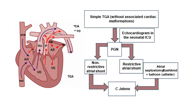
TGA is a cardiac catheterization (CHD) in which the aorta is connected to the right ventricle (RV) and the pulmonary artery to the left ventricle (LV), thus creating a parallel circulation and requiring intracardiac communications to allow survival.11 After birth, there is a decrease in pulmonary vascular resistance (PVR), leading to increased pulmonary flow and venous return to the left atrium (LA). This increased pressure in the LA can cause partial or total closure of the ductus arteriosus (FO), reducing the atrial shunt (LA→RA) and impairing oxygenation of the RV and aorta (Figure 2).12


Incidence


TGA is the most frequent cyanotic heart disease in the neonatal period, accounting for 5-7% of all congenital heart defects. A higher prevalence is observed in males, with a ratio of 2:1, in fetuses of pregnant women with pre-gestational diabetes.


Prenatal diagnosis


Diagnosis is possible through fetal echocardiography, and it is an important factor in prognosis, potentially reducing morbidity and mortality related to this CHD. It is important to note that despite technological advances, prenatal diagnosis of TGA remains low (<50%), particularly in simple TGA, i.e., with an intact interventricular septum, as the fetal heart image in the four-chamber view is normal in such cases.


Postnatal managementl


Echocardiography is essential for evaluating FO or atrial septal defect (ASD). In cases where the atrial shunt is restrictive, balloon atrial septostomy (Rashkind procedure) is performed to stabilize the newborn.13 Administration of prostaglandin E1 (PGE1) and atrial septostomy are recommended practices to maintain interatrial circulation before anatomical correction, although simple patency of the atrium is not sufficient to prevent hypoxia and acidosis.13 In certain situations, TGA may be associated with pulmonary hypertension, causing right-to-left atrial septal shunt, resulting in “paradoxical cyanosis” (pre-ductal saturation lower than post-ductal).14 In these clinical presentations, pulmonary vasodilators such as milrinone and nitric oxide (NO) are indicated to preserve cerebral oxygenation and improve hemodynamics.15 Current surgical correction of TGA – Jatene surgery (arterial switch) – should ideally be performed between 7 days and 3 weeks of life, before regression of the left ventricle with sustained drop in SVR.16 Figure 2 illustrates the postnatal management of the newborn with simple TGA (= TGA without associated malformations).


Figure 2 - Schematic drawing of the parallel circulation and flowchart illustrating the postnatal management of the newborn with simple TGA (= TGA without associated malformations). VCS: superior vena cava; IVC: inferior vena cava; AD: right atrium; VD: right ventricle; VP: pulmonary vein; Ao: aorta; AE: left atrium; VE: left ventricle; P: pulmonary artery; **FO: forame ovale; *CA: ductus arteriosus; ICU: intensive care unit; PGN: prostaglandin. C: Cardiac surgery; Jatene:  switch arterial surgery.


3. Ebstein’s anomaly


Understanding


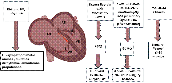
Ebstein’s anomaly is defined as the apical displacement of the tricuspid valve (TV) implantation. It results from the failure of its cusps to delaminate during cardiogenesis. Thus, the TV is not in its usual location within the right ventricle (RV), but rather displaced towards the apex of the heart. This results in a reduction of functional RV caused by “atrialization” of its inlet (Figure 3). The greater the displacement of its septal and inferior cusps, the smaller the functional RV and the greater the symptoms.17


Incidence and etiopathogenesis


It occurs in 3-7% of fetal heart defects and in 1 out of every 20,000 live births. Ebstein’s anomaly has been associated with maternal ingestion of lithium carbonate, a drug used to treat manic-depressive psychosis, suggesting that lithium may be a specific teratogen.18


Prenatal diagnosis


The fetal diagnosis of Ebstein’s anomaly is made using a four-chamber view of the fetal heart on ultrasound. Apical displacement of the true tricuspid valve annulus greater than 8 mm/m2 relative to the mitral annulus confirms the diagnosis.19 There is a risk of heart failure with cardiomegaly resulting from increased tricuspid regurgitation, tachyarrhythmia, and the appearance of fetal hydrops. Early cardiomegaly can compromise normal lung development. Neonatal and fetal death due to early circular shunt caused by ventilatory difficulties is not uncommon.


Postnatal management


It is not uncommon for neonatal fetal death to occur due to ventilatory distress in the most severe cases. Worsening intrauterine conditions may indicate the need to terminate the pregnancy before pulmonary maturity. Delivery should take place in a tertiary hospital, under the care of a pediatric cardiology and a pediatric intensive care team. Severe forms of Ebstein’s anomaly associated with decreased pulmonary blood flow, severe cyanosis, and prostaglandin E1 is indicated to maintain a patent ductus arteriosus, followed by palliative surgery (systemic-pulmonary shunt).

In less severe presentations of Ebstein’s anomaly, varying degrees of cyanosis are found, which generally decrease after the first few days as pulmonary vascular resistance falls. In presentations of Ebstein’s anomaly with congestive heart failure, amines and diuretics are indicated. Arrhythmias, when present, require antiarrhythmic treatment (amiodarone, propafenone, among others). Corrective surgery may involve valve repair, currently the most widely used technique for conical reconstruction of the tricuspid valve, as described by Dr. José Pedro da Silva’s team, and is generally performed between 12 and 18 months of age.20,21 Figure 3 illustrates postnatal management.


Figure 3 - Schematic drawing of the anatomical characteristics and flowchart illustrating the postnatal management of the newborn with Ebstein's anomaly. HF: heart failure;AD: right atrium; VD: right ventricle; AE: left atrium; VE: left ventricle; M: mitral valve; T: tricuspid valve; PGE1: prostaglandin E1; BT: Blalock-Taussig surgery (systemic-pulmonary anastomosis); Starnes:  tricuspid valve closure; “Cone”: tricuspid valve cone technique (= repositioning of the tricuspid valve). 


44. Arrhythmias


4.1. Complete Atrioventricular Block (TAVB)


Understanding


In complete or total atrioventricular block (CAVB), there is a complete dissociation between atrial and ventricular activity, with a ventricular rate usually below 60 bpm.22


Incidence


It occurs in 1 in every 15,000 to 20,000 live births, with high overall mortality, being 75-90% in CAVB associated with structural heart disease and about 20% in autoimmune AV block with a structurally normal heart. CAVB can occur in fetuses without structural heart disease or be associated with heart conditions such as left isomerism (14-42% of cases), congenitally corrected transposition of the great arteries, and unbalanced atrioventricular septal defect (AVSD). In fetuses without structural heart disease, autoimmune congenital CAVB is the most frequent. The risk for fetal CAVB in pregnant women with positive anti-Ro and/or anti-La antibodies is 1-5%, and in those with hypothyroidism the risk is 9 times higher, and the risk of recurrence is 11-19% (previous child with CAVB).23 Anticonvulsant agents, retinoic acid, and viral infections may also be related to non-autoimmune CAVB without structural disease.23


Diagnosis and prenatal management


In echocardiography, the assessment of fetal heart rate (FHR) and rhythm is performed by recording the simultaneous assessment of atrial and ventricular systole. In general, we usually use Doppler to record the inflow (mitral) and outflow (aortic) pathways of the left ventricle, where the A wave of the mitral flow represents atrial systole, and the aortic flow represents ventricular systole (V). Thus, the measurement of the time between the A and V waves reflects atrioventricular electrical conduction, and its correlation with mechanical PR has already been validated, making it possible to detect AV conduction abnormalities such as atrioventricular block.23,24

In autoimmune AV block, passive passage of autoantibodies via the placenta occurs from 11 weeks onwards, and the peak risk period for AV block is between 18-24 weeks. Thus, the Brazilian Fetal Cardiology Guideline recommends monitoring fetal heart rate (FHR) and AV interval by weekly serial fetal echocardiogram between 18-26 weeks of gestation. After 26 weeks, evaluate every 4 weeks whether the AV interval (=mechanical PR interval) remains stable or is normal, and if there is a progressive increase in this interval or if it is greater than 150ms, maintain weekly evaluation.22

In this scenario, the following are considered at high risk for developing fetal CAVB: mothers with high titers of anti-Ro/anti-SSA (> 50U/ml) and anti-Ro kD52, anti-Ro kD5260 and 48 kD La (anti-SSB) subtypes, and fetuses with increased atrial echogenicity and/or moderate or severe tricuspid regurgitation.25,26 Monitoring fetal cardiac function is crucial in fetuses with AV block.

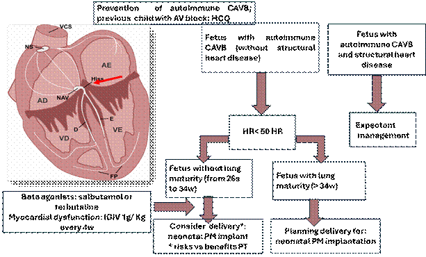
There is no guideline for evaluating fetal cardiac function as there is for anatomical evaluation, and several parameters can be used for this evaluation. The cardiovascular score, also known as the 10-point score, was validated by Prof. James Huhta and combines basic Doppler ultrasound parameters. This score is used to assess fetuses at risk for heart failure and evaluates the risk prediction for hydrops fetalis and perinatal mortality.23,27 Figure 4 illustrates the prenatal management of CAVB.


Figure 4 - Schematic drawing of the heart's electrical conduction system and flowchart illustrating the prenatal management of complete atrioventricular block (CAVB). HCQ: hydroxychloroquine; HR: heart rate; PM: pacemaker; PT: prematurity; CS: superior vena cava; NS: sinoatrial node; AD: right atrium; NAV: atrioventricular node; E: left; D: right; FP: Purkinje fibers; VD: right ventricle; AE: left atrium; VE: left ventricle; IVIG: intravenous immunoglobulin; W: weeks of gestation. .22,23,28,29,30,31


Postnatal management


A hemodynamically stable newborn with a heart rate > 50 bpm and no symptoms does not require urgent pacemaker implantation. In hydropic infants and/or those with a heart rate < 50 bpm, delivery should be planned for immediate postnatal pacemaker implantation. In the delivery room, the pediatrician should observe signs of cardiovascular compromise, such as pallor and poor perfusion: provide oxygen support, combat hypothermia, and install an ECG monitor. If TAVB is present with signs of low cardiac output: administer epinephrine 0.01 mg/kg intravenously or 0.1 mg/kg endotracheally, or atropine 0.02 mg/kg IV (minimum dose 0.1 mg and maximum 0.5 mg). Continuous infusion of epinephrine or, when available, isoproterenol, can be installed for hemodynamic support prior to cardiac pacemaker implantation.23,30,31 In cases where delivery occurred in a hospital without cardiac surgery resources, transcutaneous pacing can be a temporary alternative until the newborn is transported.32

How to install the transcutaneous pacemaker? On the same equipment used for cardioversion (CV), turn the knob (arrow) to change to pacemaker mode, set the pacemaker mode to asynchronous, set the desired heart rate to the appropriate one for age, and select the current to be used (we generally start with 5 mA, considering increasing to 10 mA), change the CV pads to adhesive pads. The adhesive pads should be in an anteroposterior position on the chest or in an infraclavicular position and cardiac apex (4 cm distance between the adhesive pads). To assess whether the pacemaker's electrical current is effective: observe the increase in heart rate and the presence of a pacemaker spike before the QRS complex on the ECG monitor.33


4.2. Tachycardias


Tachyarrhythmias can be divided into: sinus tachycardia (ST), supraventricular tachycardia (SVT), and ventricular tachycardia (VT). The most common type is SVT, accounting for approximately 70% to 90% of cases in the fetal period, atrioventricular (AV) with a risk of progression to hydrops of 30% to 40%.34,35


Supraventricular tachycardia (SVT)


Understanding


Supraventricular reentrant tachycardia is the most common type of tachycardia in the fetus and child. It is generally caused by a reentrant circuit in which the AV node conducts the impulse anterogradely from the atria to the ventricles, and a fast accessory pathway conducts the ventricular impulse back to the atria. Atrial and ventricular rates are identical, with a fetal heart rate of 220 to 300 bpm with 1:1 conduction and a VA/AV ratio < 1 (short VA interval).34,35


Incidence


Most hearts are structurally normal, but Ebstein’s anomaly is known to be associated with accessory pathways. During the Covid-19 pandemic, some studies have demonstrated an increasement in the incidence of supraventricular tachycardia, likely because of fetal hypoxia caused by SARS-CoV-2 infection in the placenta.36,37


Diagnosis and prenatal management


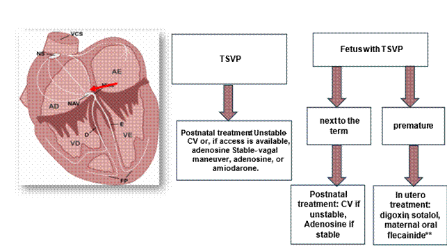
Fetal heart rate can be assessed and monitored by Doppler ultrasound and fetal echocardiography. Prenatal diagnosis of LVSD can be performed non-invasively by Doppler analysis or by analyzing the movements caused by atrial and ventricular contractions during systole. Atrial systole corresponds to the A wave in AV valve Doppler, by conventional or tissue Doppler, and in venous Doppler (pulmonary vein, vena cava, and ductus venosus) and via M-mode by atrial wall contraction. Ventricular systole is identified by Doppler of aortic or pulmonary flow, or by systolic movement of the ventricular wall in M-mode.13,22,23-25 The cardiovascular score (10-point score), which combines basic Doppler ultrasound parameters, is used to assess signs of heart failure. Prenatal management is described in the flowchart in Figure 5.


Postnatal management


In a newborn (NB) with supraventricular tachycardia (SVT), classically the heart rate (HR) will be above 220 bpm, with an absent or abnormal P wave, narrow QRS (duration < 0.09 seconds), and non-variable HR (regular R-R interval) on the ECG monitor or 12-lead ECG. The ECG should not delay management; thus, for initial management, the ECG monitor is sufficient. In SVT with hemodynamic instability (signs of low cardiac output or heart failure): oxygen support, ECG monitor, monitor blood pressure and oximetry, and synchronized electrical cardioversion at an initial charge of 0.5 to 1 J/kg, increasing to 2 J/kg in subsequent charges.

Attention to cardioversion (CV): cardiopulmonary resuscitation equipment in “standby mode”, pre-CV analgesia, and conductive gel to prevent skin burns. All these procedures should not delay CV. In the presence of venous access, adenosine can be used. In SVT, with hemodynamic stability (without signs of low cardiac output or heart failure): oxygen support, ECG monitor, monitor blood pressure and oximetry, vagal maneuver (ice on the face) and if the vagal maneuver fails, reverse with adenosine intravenously (or even intraosseous). Administer adenosine with 5ml of saline solution as a bolus at a dose of 0.1mg/kg/dose and if necessary, repeat with double the first dose (maximum doses: 6mg in the 1st dose and 12mg in the subsequent dose).

Attention: all cardiopulmonary resuscitation equipment must be prepared and checked before using adenosine. After reversal of SVT, antiarrhythmic medication with beta-blocker or amiodarone should be discussed with a cardiologist.30 Figure 5 illustrates postnatal management in SVT.


Figure 5 - Schematic drawing of the heart's electrical conduction system and flowchart illustrating the prenatal management of supraventricular tachycardia. SVT: supraventricular tachycardia; VCS: superior vena cava; NS: sinoatrial node; AD: right atrium; NAV: atrioventricular node; LE: left bundle of His; R: right bundle of His; FP: Purkinje fibers; VD: right ventricle; AE: left atrium; VE: left ventricle; CV: electrical cardioversion.


Planning the delivery of fetuses diagnosed with congenital heart disease

Birth planning should consider three main factors: [1] the risk of hemodynamic instability at birth; [2] the resources of the region; [3] the presence of obstetric complications.


Choosing a hospital and transporting your newborn

Most newborns with CHD do not require specialized perinatal care, and recommendations are made for them to be born at the local hospital and followed up as outpatients.16 However, if the presence of a specialized pediatric cardiology team and postpartum intensive care is required, the place of birth should take these special needs into account, avoiding the deleterious effects of transport on these newborns.6  Transporting these critically ill newborns requires mobile ICU units with a specialized healthcare team, infusion pumps for medications such as prostaglandins and amines, ventilatory support, and saturation and cardiac monitors.


Gestational age for delivery

Recent studies have shown that in fetuses diagnosed with severe CHD, delivery tends to occur earlier than in those in which diagnosis of CHD is made after birth.17 This finding is particularly concerning, given that healthy newborns born between 37 and 38 weeks have a higher risk of worse outcomes compared to those born later between 39 and 40 weeks.6,18 Studies have shown that newborns with CHD have a longer stay in the intensive care unit and higher mortality when born before 39 weeks.18 Therefore, in the absence of fetal or maternal indications for prematurity, the potential advantages of elective preterm delivery of fetuses with CHD should be carefully considered.6

In addition to increased mortality, there is increasing evidence that the decision on the timing of delivery of fetuses with CHD should also consider the potential effect of gestational age at birth on neurological outcome and, therefore, delivery of these babies at term or as close to term as possible may improve brain development and decrease susceptibility to postnatal injury.6


Delivery method

Most experts agree that, in the absence of heart failure, hemodynamic decompensation, fetal hydrops, or sustained fetal arrhythmia, elective preterm delivery offers no advantage.6 It is frequently stated that normal vaginal delivery should be the goal in fetal CHD,6 reserving cesarean section for obstetric indications, with which we agree.

Data from retrospective studies show that prenatal diagnosis of severe CHD, such as HLHS, TGA, double outlet right ventricle, or tetralogy of Fallot, increases the likelihood of elective delivery and cesarean section. In fetuses with CHD, the mode of delivery has not been shown to affect the Apgar score, pre- and post-surgical morbidity, including the risk of hemodynamic instability, metabolic acidosis and end-organ dysfunction, length of hospitalization, or survival to surgery or hospital discharge.

Two retrospective studies concluded that labor is safe for fetuses with CHD in most cases,19 but the impact on long-term functional outcomes and neurological development is largely unknown.6


Fetal vitality during labor

The decision to perform a vaginal delivery in women with a prenatal diagnosis of fetal CHD opens a debate about how to monitor these fetuses during labor, with the aim of promptly identifying and intervening for those at risk of hypoxemia and minimizing the risk of hypoxic-ischemic encephalopathy and adverse long-term neurological outcomes.

Some retrospective studies evaluating the use of cardiotocography during labor in fetuses with CHD have shown that these fetuses have a higher percentage of non-reassuring tracings, but no characteristic pattern of fetal heart rate has been related to specific cardiac pathologies. As in normal fetuses, the use of continuous CTG in labor in fetuses with CHD has been associated with an increased rate of emergency cesarean delivery.20


CONCLUSIONS

Intrauterine detection of CHD allows for better prenatal counseling and delivery planning, especially when the need for urgent postnatal intervention is anticipated based on available predictive models. Perinatal management should be tailored to the specific needs of the mother and fetus, and should include decisions regarding location, timing, and mode of delivery that generally minimize the risk of premature or operative delivery. In selected cases, there may be maternal or fetal indications for early delivery, including a variety of obstetric indications such as spontaneous onset of labor, maternal comorbidities, pregnancy complications, or non-reassuring results from fetal viability tests.

Collaboration between specialized obstetric and pediatric services and careful attention to perinatal management and delivery planning after a prenatal diagnosis of CHD can improve the perinatal status of newborns with the potential for improvement in both survival and long-term outcomes.


REFERENCES


1.      Haxel CS, Johnson JN, Hintz S, Renno MS, Ruano R, Zyblewski SC, et al. Care of the Fetus with Congenital Cardiovascular Disease: From Diagnosis to Delivery. Pediatrics. 2022 ;150 (Suppl 2): e2022056415C. doi: 10.1542/peds.2022-056415C.

2.      Mehta LS, Warnes CA, Bradley E, Burton T, Economy K, Mehran R, et al. American Heart Association Council on Clinical Cardiology; Council on Arteriosclerosis, Thrombosis and Vascular Biology; Council on Cardiovascular and Stroke Nursing; and Stroke Council. Cardiovascular Considerations in Caring for Pregnant Patients: A Scientific Statement from the American Heart Association. Circulation. 2020;141(23):e884.

3.      Donofrio MT, Skurow-Todd K, Berger JT, McCarter R, Fulgium A, Krishnan A, Sable CA. Risk-stratified postnatal care of newborns with congenital heart disease determined by fetal echocardiography. J Am Soc Echocardiogr. 2015;28(11):1339- 49.doi 10.1016/j.echo2015.07.005.

4.      Vieira MF, BravoValenzuela NJ, Júnior EA. ZScore in Fetal Echocardiography – Is there Still Room for New Studies? Congenit Heart Dis. 2024;19(3):305-14. https://doi.org/10.32604/chd.2024.053484

5.      Herdy GV; Silva AE; Lucas E; Bravo-Valenzuela NM. Cardiologia Pediátrica: Prática Clínica. 1. ed. Rio de Janeiro: Thieme Revinter, 2021:169-174.

6.      Grossfeld P, Nie S, Lin L, Wang L, Anderson RH. Hypoplastic Left Heart Syndrome: A New Paradigm for an Old Disease? J Cardiovasc Dev Dis. 2019;6(1):10. doi: 10.3390/jcdd6010010.

7.      Yabrodi M, Mastropietro CW. Hypoplastic left heart syndrome: from comfort care to long-term survival. Pediatr Res. 2017;81(1-2):142-9.

8.      Silva JP; Lopes LM; Silva LF. Síndrome do coração esquerdo hipoplásico. In: Crotti UA, Mattos SS, Pinto JR. Cardiologia e cirurgia cardiovascular pediátrica. 2ª ed. São Paulo: Roca; 2012: 637-60.

9.      Park MK. Cyanotic congenital heart defects.In: Park MK, Salamat M. Pediatric cardiology for practioners. 7th ed. Philadelphia: Elsevier:2021: 1275-88.

10.  Chen JM, Ittenbach RF, Lawrence KM, Hunt ML, Kaplinski M, Mahle M, et al. Increased utilization of the hybrid procedure is not associated with improved early survival for newborns with hypoplastic left heart syndrome: a single-centre experience. Eur J Cardiothorac Surg. 2024;65(4):ezae164. doi: 10.1093/ejcts/ezae164.

11.  Herdy GV; Silva AE; Lucas E; Bravo-Valenzuela NM. Cardiologia Pediátrica: Prática Clínica. 1. ed. Rio de Janeiro: Thieme Revinter, 2021:119-125.

12.  Leite MF; Silva AE. Cardiologia pediátrica. Série Pediatria Soperj. 2ª ed. Baueri: Manole; 2019: 227-37.

13.  Subramanian S, Jani S, Well A, Mikulski MF, Agrawal H, Byron Holt D, et al. Revisiting the Role of Balloon Atrial Septostomy Prior to the Arterial Switch Operation. World J Pediatr Congenit Heart Surg. 2024;15(6):746-52. doi: 10.1177/21501351241252428

14.  Park MK. Cyanotic Congenital Heart Defects. In: Park MK, Salamart M. Park’s Pediatric Cardiology for Practitioners, 7th ed. Philadelphia: Elsevier; 214-7.

15.  Leite MF; Silva AE. Cardiologia pediátrica. Série Pediatria Soperj. 2ª ed. Baueri: Manole; 2019: 227-37.

16.  Donofrio MT, Moon-Grady AJ, Hornberger LK, Copel JA, Sklansky MS, Abuhamad A et al; Diagnosis and treatment of fetal cardiac disease: a scientific statement from Aerican Heart Association. Circulation 2019: 27; 129(21); 2183-242.

17.  Anderson RH. Understanding Ebstein’s malformation. Cardiol Young. 2015;25(1):137-8.

18.  G.K. Sharland, S.K. Chita, L.D. Allan.  Tricuspid valve dysplasia or displacement in intrauterine life J Am Coll Cardiol. 1991; 17(4):944-9. doi: 10.1016/0735-1097(91)90877-c.

19.  Farias CVB. Anomalia de Ebstein e displasia da valva tricúspide. In: Bravo-Valenzuela NJM, Lucas E, Silva AEA, Farias CVB. Atlas de ecocardiografia fetal. Rio de Janeiro: Thieme Revinter Publicações, 2021. p. 151-7.

20.  Sainathan S, da Fonseca da Silva L, da Silva JP. Ebstein’s anomaly: contemporary management strategies. J Thorac Dis 2020;12(3):1161-73.

21.  Silva JP, Silva L da F, Moreira LF, Lopez LM, Franchi SM, Lianza AC, et al. Cone reconstruction in Ebstein’s anomaly repair: early and long-term results. Arq Bras Cardiol 2011;97(3):199-208. doi: 10.1590/s0066-782x2011005000084.

22.  Pedra SRFF, Zielinsky P, Binotto CN, Martins CN, Fonseca ESVBD, Guimarães ICB, et al. Brazilian Fetal Cardiology Guidelines - 2019. Arq Bras Cardiol. 2019; 112(5):600-48.

23.  Donofrio MT, Moon-Grady AJ, Hornberger LK, Copel JA, Sklansky MS, Abuhamad A, et al. American Heart Association Adults with Congenital Heart Disease Joint Committee of the Council on Cardiovascular Disease in the Young and Council on Clinical Cardiology, Council on Cardiovascular Surgery and Anesthesia, and Council on Cardiovascular and Stroke Nursing. Diagnosis and treatment of fetal cardiac disease: a scientific statement from the American Heart Association. Circulation. 2014;129(21):2183-242. doi: 10.1161/01.cir.0000437597.44550.5d. Erratum in: Circulation. 2014 May 27;129(21):e512.

24.  Glickstein J, Buyon J, Kim M, Friedman D; PRIDE investigators. The fetal Doppler mechanical PR interval: a validation study. Fetal Diagn Ther. 2004;19(1):31-4. doi: 10.1159/000074256.

25.  25.Hunter LE, Simpson JM. Atrioventricular block during fetal life. J Saudi Heart Assoc. 2015; 27(3):164-78. doi: 10.1016/j.jsha.2014.07.001.

26.  Kashou AH, Goyal A, Nguyen T, et al. Atrioventricular Block. [Updated 2024 Feb 12]. In: StatPearls [Internet]. Treasure Island (FL): StatPearls Publishing; 2025. Available from: https://www.ncbi.nlm.nih.gov/books/NBK459147.

27.  Huhta JC. Fetal congestive heart failure. Semin Fetal Neonatal Med. 2005; 10(6):542-52.

28.  Götestam Skorpen C, Hoeltzenbein M, Tincani A, Fischer-Betz R, Elefant E, Chambers C, et al. The EULAR points to consider for use of antirheumatic drugs before pregnancy, and during pregnancy and lactation. Ann Rheum Dis. 2016;75(5):795-810. doi: 10.1136/annrheumdis-2015-208840.

29.  Groves AM, Allan LD, Rosenthal E. Therapeutic trial of sympathomimetics in three cases of complete heart block in the fetus. Circulation. 1995; 92(12):3394-6. doi: 10.1161/01.cir.92.12.3394.

30.  Batra AS, Silka MJ, Borquez A, Cuneo B, Dechert B, Jaeggi E, et al. Pharmacological Management of Cardiac Arrhythmias in the fetal and Neonatal Periods: A Scientific Statement From the American Heart Association: Endorsed by the Pediatric & Congenital Electrophysiology Society (PACES). Circulation. 2024; 149(10): e937 -e952. doi: 10.1161/CIR.0000000000001206.

31.  Pruetz JD, Miller JC, Loeb GE, Silka MJ, Bar-Cohen Y, Chmait RH. Prenatal diagnosis and management of congenital complete heart block. Birth Defects Res. 2019;111(8):380-8. doi: 10.1002/bdr2.1459.

32.  Aziz K, Lee HC, Escobedo MB, Hoover AV, Kamath-Rayne BD, Kapadia VS, et al. Part 5: Neonatal Resuscitation: 2020 American Heart Association Guidelines for Cardiopulmonary Resuscitation and Emergency Cardiovascular Care. Circulation. 2020;142 (16_suppl_2): S524-S550. doi: 10.1161/CIR.0000000000000902.

33.  starship.org.nz [Internet]. New Zealand: Starship Hospital. Pacemakers (temporary) for a child [cited 2025 Aug 13]. Available from: [https://starship.org.nz/guidelines/pacemaker-temporary-for-a-child/].

34.  Bravo-Valenzuela NJ, Rocha LA, Machado Nardozza LM, Araujo Júnior E. Fetal cardiac arrhythmias: Current evidence. Ann Pediatr Cardiol. 2018; 11(2):148-63.

35.  Tang J, Huang P, Deng X, Zhao L, Zhai Y, Wang T. Advances and challenges of prenatal interventions for fetal tachyarrhythmias. Front Pediatr. 2024; 12:1509158. doi: 10.3389/fped.2024.1509158.

36.  Samples S, Patel S, Lee S, Gotteiner N, Patel A. Incidence of fetal arrhythmia before and during the COVID-19 pandemic: a single-center experience. Pediatr Cardiol. 2025;46(2):431-6. doi: 10.1007/s00246-024-03439-3.

37.  Mullins J, Bewley DJ, Oviedo A. COVID-19 and placental infection: are fetal survivors at risk of long-term cardiovascular complications? Cureus. 2023; 15(4):e38077. doi: 10.7759/cureus.38077.


Scientific Editor:

Fernanda Pinto Mariz

ORCID: https://orcid.org/0000-0002-6981-2352


Editor:

Sociedade de Pediatria do Rio de Janeiro – SOPERJ

E-mail: secretaria@soperj.org.br


Financial support:

None.


Availability of research data:

the underlying content of the research text is contained in the article.


Conflict of interests:

None.


Authors’ contributions:

NJM Bravo-Valenzuela: data collection, conceptualization, project management, methodology, drafting - preparation of the original manuscript, drafting - revision and editing, supervision.

E Lucas: data collection, conceptualization, resource management, project management, methodology, drafting - preparation of the original manuscript, drafting - revision and editing, supervision, visualization.

RP Correia: drafting - preparation of the original manuscript, visualization.


Rev Pediatria SOPERJ 2026;26(2): e20260375