Flowchart 1. Selection of references
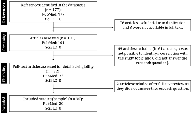
n = number of studies.
REVIEW ARTICLE
Submitted: 25/08/2025
Approved: 01/10/2025
DOI: http://dx.doi.org/10.31365/issn.2595-1769.2026.0386
Literature Review: Arrythmias in Pediatric Patients with Kawasaki Disease
Revisão de Literatura: Arritmias em Pacientes Pediátricos com Doença de Kawasaki
Revisión de la Literatura: Arritmias en Pacientes Pediátricos con Enfermedad de Kawasaki
Maria Fernanda Trepin Granato Acciarito1,2
Gustavo Rodrigues Prado1,3
Vinicius Rodrigues Prado1,4
Camila Laurindo e Silva1,5
Luciano Rodrigues Costa1,6
1 Centro Universitário de Volta Redonda, Pediatria. Volta Redonda-RJ, Brasil.
2ORCID: https://orcid.org/0000-0002-0978-103X
3ORCID: https://orcid.org/0000-0001-8208-169X
4 ORCID: https://orcid.org/0000-0001-6287-8554
5ORCID: https://orcid.org/0000-0003-2190-1417
6ORCID: https://orcid.org/0000-0001-8657-2656
Autor correspondente:
Maria Fernanda Trepin Granato Acciarito1
E-mail: mariafernanda_granato@hotmail.com
Abstract
Introduction: This literature review was conducted to identify the most frequent arrhythmias in children and adolescents with Kawasaki disease, at each phase of the disease, to assist teams in identifying them appropriately. It is known that this vasculitis is the leading cause of acquired cardiovascular disease in developed countries, thus, there is importance in expanding knowledge about it. Objective: In this article, gather all reports of arrhythmias related to Kawasaki Disease. Data sources: PubMed and SciELO databases were used, using MeSH descriptors "Mucocutaneous Lymph Node Syndrome," "Cardiovascular Disease," and "Arrhythmias". We found 177 papers and selected 30. Papers that did not answer the question, duplicates, or those not available in full were excluded. We analyzed papers from 1980 to 2023. Data synthesis: 105 patients diagnosed with Kawasaki disease, from case reports, retrospective studies, or cohorts, presented arrhythmias. They were divided into three groups according to the phase of the disease - acute, subacute and chronic - based on case reports. Conclusions: The most common arrhythmia in the acute and chronic phases was sinus tachycardia. In addition to this, in the chronic phase, premature ventricular contractions and right bundle branch block were also more frequent.
Keywords: Kawasaki Disease; Arrhythmia; Cardiovascular Disease.
RESUMO
Introdução: Esta revisão da literatura foi feita visando identificar as arritmias mais frequentes em crianças e adolescentes com doença de Kawasaki, em cada fase da doença, a fim de auxiliar as equipes a identificar da maneira adequada. Sabe-se que esta vasculite é a maior causa de doença cardiovascular adquirida em países desenvolvidos, daí a importância em ampliar os conhecimentos sobre ela. Objetivo: Reunir neste artigo todos os relatos de arritmias relacionados à doença de Kawasaki. Fontes dos dados: Foram utilizadas as bases de dados PubMed e SciELO, utilizando descritores do MeSH “Mucocutaneous Lymph Node Syndrome”, “Cardiovascular Disease” e “Arrhythmias”. Foram encontrados 177 trabalhos, dos quais 30 foram selecionados. Os trabalhos que não responderam à pergunta, os que estavam duplicados ou não disponíveis na íntegra foram excluídos. Analisamos trabalhos de 1980 a 2023. Síntese dos dados: 105 pacientes com diagnóstico de doença de Kawasaki, sejam estes advindos de relatos de caso, estudos retrospectivos ou coortes, apresentaram arritmias. Foi feita uma divisão em três grupos, de acordo com a fase da doença - aguda, subaguda e crônica -, com base nos relatos de casos. Conclusões: A arritmia mais comum na fase aguda e crônica foi a taquicardia sinusal. Além desta, na fase crônica, contrações ventriculares prematuras e bloqueio de ramo direito também foram mais frequentes.
Palavras-chave: Síndrome de Linfonodos Mucocutâneos; Arritmias Cardíacas; Doenças Cardiovasculares.
Resumen
Introducción: Esta revisión de la literatura tuvo como objetivo identificar las arritmias más frecuentes en niños y adolescentes con enfermedad de Kawasaki en cada etapa de la enfermedad, con el fin de ayudar a los equipos a identificarlas adecuadamente. Se sabe que esta vasculitis es la principal causa de enfermedad cardiovascular adquirida en países desarrollados, de ahí la importancia de ampliar el conocimiento sobre ella. Objetivo: Recopilar en este artículo todos los reportes de arritmias relacionadas con la enfermedad de Kawasaki. Fuentes de datos: Se utilizaron las bases de datos PubMed y SciELO, empleando los descriptores MeSH “Mucocutaneous Lymph Node Syndrome”, “Cardiovascular Disease” y “Arrhythmias”. Se encontraron 177 estudios, de los cuales 30 fueron seleccionados. Se excluyeron los estudios que no respondieron la pregunta, aquellos que eran duplicados o aquellos no disponibles en su totalidad. Analizamos estudios de 1980 a 2023. Síntesis de datos: 105 pacientes diagnosticados con enfermedad de Kawasaki, ya sea de informes de casos, estudios retrospectivos o cohortes, presentaron arritmias. Se dividieron en tres grupos según la fase de la enfermedad (aguda, subaguda y crónica), según informes de casos. Conclusiones: La arritmia más frecuente en las fases aguda y crónica fue la taquicardia sinusal. Además, las extrasístoles ventriculares y el bloqueo de rama derecha del haz de His también fueron más frecuentes en la fase crónica.
Palabras clave: Síndrome del Ganglio Linfático Mucocutáneo; Arritmias Cardíacas; Enfermedades Cardiovasculares.
Kawasaki disease (KD) is an acute, febrile, self-limiting systemic vasculitis observed in early childhood, most commonly in children under 5 years of age, accounting for approximately 80% of cases.1,2 It is currently the second most common vasculitis in pediatrics, after Henoch-Schönlein purpura. The disease was first described in Japan in 1967 by Tomisaku Kawasaki.3,4 It is now known to have a worldwide distribution, although it predominates in Asia and in children of Asian descent. It is considered the leading cause of acquired heart disease in industrialized countries.4,5
There are no specific biomarkers for the diagnosis of Kawasaki disease; therefore, the diagnostic criteria for typical Kawasaki are clinical and include fever lasting five days, in addition to four of the following five clinical features: oropharyngeal changes; non-purulent conjunctivitis; acute cervical lymphadenopathy with lymph node diameter > 1.5 cm; Peripheral changes in the extremities; or a generalized polymorphic rash.2,6
However, there are cases in which all criteria are not met, which can delay diagnosis if the possibility of incomplete Kawasaki disease is not raised. In this scenario, we should consider incomplete Kawasaki disease in children with fever for five or more days and at least two of the clinical criteria of the complete form of the disease, or fever for seven or more days without another explanation for the condition. Therefore, laboratory tests should be performed to look for findings that may suggest the disease, such as elevated C-reactive protein levels ≥ 3.0 mg/dL and/or erythrocyte sedimentation rate ≥ 40 mm/h. If the child presents with abnormalities in these tests, the investigation should continue with an echocardiogram, looking for typical findings, and complementary laboratory tests. Three or more of the following findings support the diagnosis: albumin level ≤ 3.0 g/dL, anemia for age, platelet count ≥ 450,000 after the seventh day of fever, white blood cell count ≥ 15,000 mm³, and urinary white blood cell count ≥ 10 per visual field.7
In Kawasaki disease, the coronary arteries are most affected, which can lead to asymptomatic coronary artery ectasia or the formation of an aneurysm.8 The development of coronary lesions occurs in 15 to 25% of untreated cases of the disease. These patients have a high risk of developing myocardial ischemia or suffering sudden death. And some sudden cardiac deaths after KD may be related to arrhythmias.3,4
During the clinical course, careful monitoring of cardiovascular function and coronary artery diameter is necessary, but, in addition to these, given that arrhythmias can lead to catastrophic outcomes, electrocardiographic rhythm monitoring is important along with the other measures.
DATA COLLECTION AND SYNTHESIS
We conducted a systematic review using searches in three databases: PubMed (U.S. National Library of Medicine), SciELO (Scientific Electronic Library Online), and BVS (Virtual Health Library), using the English MeSH descriptors “Mucocutaneous Lymph Node Syndrome”, “Cardiovascular Disease”, and “Arrhythmias”. The 2020 PRISMA methodology was used for the selection and organization of the analyzed studies, according to Flowchart 1. In total, 177 studies were found, of which 30 were included in the sample, among them case reports, retrospective studies, and cohorts. No inclusion criterion was included for year of publication, aiming to broaden the sample size. Therefore, studies from 1980 to 2023 were reviewed.
Flowchart 1. Selection of references
n = number of studies.
DISCUSSION AND CONCLUSIONS
The clinical course of Kawasaki disease is described as triphasic. The acute phase of the disease lasts approximately 11 days. During this phase, pathological changes occur in small blood vessels, including perivasculitis and vasculitis. In addition, inflammation of the intima layer of medium and large arteries may occur. For the most part, clinically significant vascular pathology during the acute phase is limited to coronary artery involvement. The most common cause of death during the acute phase of the disease is myocarditis.9
The subacute phase extends from the 11th to the 20th day. During this period, fever, rash, and lymphadenopathy may disappear. Desquamation, along with arthralgia and arthritis, occurs more frequently during this phase. Cardiovascular changes during the subacute phase include coronary artery aneurysm formation, perivasculitis, and vessel wall edema. Acute myocardial infarction (AMI)
occurs most commonly during this phase, usually due to thrombosis. Although aneurysms may eventually resolve, arterial stenosis can persist indefinitely.9
During the convalescent period, which occurs from the 21st to the 60th day, most clinical features disappear. Although vascular inflammation decreases, MI can still occur. The chronic phase, which begins around day 61, can last indefinitely. Throughout the chronic phase, scarring and thickening of the intima of the coronary arteries can continue, resulting in ischemic heart disease.9
Table 1 below was compiled from the results found in our research, based on reports from 105 children and adolescents with arrhythmias caused by Kawasaki disease.
Table 1 Electrocardiographic abnormalities found
|
Arrhythmia |
Acute phase |
Subacute phase |
Chronic phase |
|
Sinus tachycardia |
8 |
- |
27 |
|
PVC |
2 |
- |
14 |
|
AVB 1st and 2nd grade Ventricular Extrasystole Right bundle branch block Ventricular tachycardia SVT Ventricular fibrillation CAVB PAC Wolff-Parkinson-White |
2 1 2 1 - - - - - |
- -
1 1 1 - - - |
4 - 27 5 1 5 1 2 1 |
This table presents the rhythms found and the number of patients described in our references.
PVC., Premature Ventricular Contraction.
AVB., Atrioventricular Block
SVT., Supraventricular tachycardia
CAVB., Complete Atrioventricular Block
PAC., Premature Atrial Contraction
Based on cardiological studies, in the acute phase of Kawasaki disease, the electrocardiogram can demonstrate any type of arrhythmias, including sinus and atrioventricular node abnormalities, with PR interval prolongation and nonspecific ST interval changes and T wave changes, generally if there is myocardial or pericardial involvement. QT interval prolongation may occur, and ventricular repolarization abnormalities have been reported. On rare occasions, malignant ventricular arrhythmias have been observed in the presence of myocarditis or myocardial ischemia.4
During the acute phase, aneurysm formation is associated with the risk of coronary thrombosis and stenosis, due to altered flow in the affected coronary arteries. In the long term, it is believed that changes in the architecture of the coronary artery walls result in endothelial cell dysfunction and potentially increase the rates of fatal and non-fatal cardiovascular events.10 There is evidence that endothelial function remains abnormal despite the resolution of aneurysms in patients with KD with aneurysm formation.11
Sinus node dysfunction and atrioventricular block are sometimes associated with inferior wall infarction caused by right coronary artery obstruction. This dysfunction and block are believed to be caused by ischemia of the sinus node and atrioventricular node. Furthermore, it was observed that the incidence of abnormal sinus node and atrioventricular function in patients with KD is apparently higher than in the normal population.3
A case report presented findings that corroborate those described by Naokata's study,3 in which a healthy 16-month-old girl presented with fever and all the classic clinical criteria for KD, and the initial electrocardiogram (ECG) revealed first-degree atrioventricular (AV) block and an incomplete right bundle branch block pattern.12
In a retrospective study conducted in a hospital in Mexico, 28 patients had arrhythmias (5.5% of the patients studied), 26 patients presented with sinus tachycardia unrelated to fever, one patient with monomorphic ventricular extrasystoles, and one patient with first-degree AV block.13
Atrioventricular block (AVB) occurs in only 0.34% of patients with Kawasaki disease, and its potential mechanisms may be attributed to inflammatory cascades and edema of the atrioventricular node. Sixty-seven percent of cases resolve spontaneously, and their average duration is 3.3 days; if AV block persists for more than a week, temporary intervention will be necessary.14
Autopsy findings in patients with Kawasaki disease have shown that lesions of the atrioventricular conduction system are classified according to the time since the onset of vasculitis. Within nine days, infiltration of inflammatory cells and edema are the main findings; then, between three and four weeks, compression of conduction cells due to perivascular edema and cellular infiltration is the main finding. From seven weeks to seven months, perivascular fibrosis and fatty infiltration are observed.3,15 Prolongation of the PR interval can be a sensitive indicator of acute inflammation of the atrioventricular conduction system.3
According to Naokata,3 40 children were evaluated; one patient was admitted with intense chest pain, pallor, and arrhythmia at 11 years of age, and the electrocardiogram showed a second-degree AVB, but in the chronic phase of the disease. This same team subjected the patients to the exercise stress test, and two patients presented with arrhythmias, premature ventricular contraction, and monomorphic ventricular tachycardia. This patient was referred to the team for recurrent palpitations at 13 years of age and had suffered from KD since 6 months of age. Selective coronary angiography showed a giant aneurysm in segment 1 and 90% stenosis in segment 6.3
The formation of coronary aneurysms in Kawasaki disease is a direct result of the inflammatory process. On days 7 to 9 of the disease, neutrophil infiltration occurs in the walls of the coronary arteries, which is rapidly replaced by monocytes, lymphocytes, and IgA plasma cells. This response causes fragmentation of the elastic lamina and damage to the media layer, resulting in the formation of aneurysms.16 As acute inflammation subsides, these remodeling areas may undergo fibrosis and form scars.17,18 Risk factors that increase a patient's risk of developing an aneurysm include delayed administration of intravenous immunoglobulin (IVIG) after the 10th day of illness,17,19 persistent fever after IVIG administration, elevated inflammatory markers, white blood cell count greater than 12,000/mL, anemia, hyponatremia, thrombocytopenia, hypoalbuminemia, male sex, and children under 12 months of age.17
It is also known that children with KD have significantly greater QTc interval dispersion in 12 leads. And increased QT interval dispersion has been associated with an increased risk of ventricular arrhythmias and sudden cardiac events.20
QT interval dispersion was assessed in 20 children from northern India with Kawasaki disease without coronary artery anomalies on echocardiography, comparing them with matched controls. Dispersion is indicative of non-homogeneous ventricular repolarization and may represent an increased risk of developing ventricular arrhythmia in this population.20
A recent study investigating the long-term prognosis of patients with coronary artery disease showed a prolonged Tpeak-Tend (Tp-e) interval in cases with sudden cardiac death compared to a group without accident. Thus, analysis of the ventricular repolarization interval and assessment of repolarization based on the Tp-e/QT ratio are of interest. The Tp-e and Tp-e/QT intervals are currently considered indicators of variation in total repolarization because the Tp-e interval includes repolarization at spatially different locations, and not just the local potential. The study also highlights that Tp-e/QT values had significant positive correlations with body temperature, which is an important point, since Kawasaki disease is associated with fever.21
In the pediatric age group, ischemic heart disease is an extremely rare condition, and this vasculitis is one of the most common underlying causes.3 Coronary artery obstruction after KD can cause MI or sudden cardiac death,3,22 and some of the sudden cardiac deaths after the disease may be related to arrhythmias. Ventricular arrhythmias have also been reported in the final stage of the disease. However, the precise mechanism of sudden death remains unknown.3
Fatal arrhythmias are suspected to be late complications of myocardial infarction. And large aneurysms frequently cause acute myocardial infarction during the first year after the onset of Kawasaki disease.23
Ventricular tachycardia (VT) may occasionally present in long-term follow-up patients with compromised left ventricular function and aneurysms, including large coronary aneurysm, coronary artery stenosis, and thrombosis.24 The mechanism of VT is believed to be reentry in patients with old AMI and automaticity or triggered activity in those with acute AMI.3
Chabali24 reports eight patients in the acute phase with sinus tachycardia correlated with fever, and that, upon cessation of the fever, the rhythm returned to sinus.
Finally, other arrhythmias reported in the acute phase were premature ventricular contraction,23,25 right bundle branch block,25 and sustained ventricular tachycardia.23 However, more records on arrhythmias in patients in the acute phase are needed, as only five studies were found.
In the subacute phase, two reports showed supraventricular tachycardia related to fever,2 ventricular tachycardia and ventricular fibrillation correlated with acute ischemia.17 In the chronic phase, other authors reported arrhythmias, which were correlated with aneurysms and ischemia, except for the case of Jeffrey et al., a supraventricular tachycardia, which had ischemic changes and an aneurysm. Ventricular tachycardia,23,28 ventricular fibrillation,13,29,30 complete atrioventricular block14 and premature ventricular contraction were also described.23,28
Shizuhiro30 reports other electrocardiographic changes in patients diagnosed with Kawasaki disease, without specifying the relationship with myocardial ischemia and the presence of aneurysms. These are: right bundle branch block, premature ventricular contraction, premature atrial contraction, Wolff-Parkinson-White syndrome and atrioventricular block.
Therefore, although arrhythmias caused by KD are not common, it is necessary for teams to be prepared to identify and treat them, avoiding unfavorable outcomes. Furthermore, this study aims to reinforce the importance of early introduction of intravenous immunoglobulin to reduce the occurrence of aneurysms,19 which are related to the triggering of arrhythmias in many of the cases reported above.
REFERENCES
1. Hicks RV, Melish ME. Kawasaki Syndrome. Pediatric Clinics of North America. 1986; 33(5):1151-75.
2. Chou CP, Lin IC, Kuo KC. A male infant had subdural effusion and paroxysmal supraventricular tachycardia during the febrile episode of Kawasaki disease: a case report and literature review. BMC Pediatrics (London). 2016; 16,71:1-6.
3. Naokata S, Kensuke K, Kazuo T, Rie I, Junji F, Osamu A, Michio M, Hiroshi K, Mamoru A, Kensuke H. Association of Sinus Node Dysfunction, Atrioventricular Node Conduction Abnormality and Ventricular Arrhythmia in Patients with Kawasaki Disease and Coronary Involvement. Circ J (Japão). 2008; 72:274-280.
4. Luis MGG, Rocío APJ, Marco AYN. Manifestaciones cardiacas en la etapa aguda de la enfermedad de Kawasaki en un hospital pediátrico de tercer nivel en la Ciudad de México. Arch Cardiol Mex (México). 2018; 88(5):441-446.
5. Yale SH, Tekiner H, Yale ES. Tomisaku Kawasaki and Kawasaki disease. Childs Nerv Syst. 2022; 38(2):233-5.
6. Rajasekaran K, Duraiyarasan S, Adefuye M, Manjunatha N, Ganduri V. Kawasaki Disease and Coronary Artery Involvement: A Narrative Review. Cureus. 2022;14(8):e28358.
7. McCrindle B, Rowley A, Newburger J, Burns J, Bolger A, Gewitz M, Baker A, Jackson M, Takahashi M, Shah P, Kobayashi T, Wu M, Saji T, Pahl E. Diagnosis, Treatment, and Long-Term Management of Kawasaki Disease: A Scientific Statement for Health Professionals From the American Heart Association. Circulation. 2017; 135(17):e927-999.
8. Crean A, Benson L, Shah A, Han K, Lesser J, McCrindle BW. Imaging the delayed complications of childhood Kawasaki disease. F1000Res. 2022; 11:147.
9. Jeffrey D, Seymour MD, Edward T, Dickinson MD. Delayed Cardiovascular Sequelae from Kawasaki Syndrome. American Journal of Emergency Medicine (Philadelphia). 1998; 16: 6.
10. Daniels LB, Gordon JB, Burns JC. Kawasaki disease: late cardiovascular sequelae. Current Opinion in Cardiology. 2012; 27(6):572-7.
11. Holve TJ, Patel A, Chau Q, Marks AR, Meadows A, Zaroff JG. Long-Term Cardiovascular Outcomes in Survivors of Kawasaki Disease. Pediatrics (Elk Groove Village 2014; 133,2: 305-311.
12. Almaleh D, Fournier A, Montigny M, Dahdah N. Rate Dependent QS Pattern in a Acute Kawasaki Disease. Congenit Heart Disease (Houston). 2010; 5: 458-461.
13. Madrid AH, Huerta EM, Serrano MC, Barcia F, Villagrá F, Rayo I. Aborted sudden death in the chronic phase of Kawasaki disease. American Heart Journal (Madrid). 1991; 121,5: 1548-1549.
14. Liu FF, Wang JJ, Wu Y, Liu HH, Hu P. Complete Atrioventricular Block and Macrophage Activation Syndrome Simultaneously Occurred in a 6-Year-Old Chinese Girl with Kawasaki Disease. Journal of Clinical Rheumatology (Hefei). 2019; 00,00: 1-2.
15. Qiu Y, Zhang Y, Li Y, Hua Y, Zhang Y. Molecular mechanisms of endothelial dysfunction in Kawasaki-disease-associated vasculitis. Front Cardiovasc. 2022; 9:981010.
16. Dietz SM, Tacke CEA, Hutten BA, Kuijpers TW. Peripheral Endothelial (Dys)Function, Arterial Stiffness and Carotid Intima-Media Thickness in Patients after Kawasaki Disease: A Systematic Review and Metanalisis. PLoS ONE. 2015;10(7):e0130913.
17. Gern BH, Mehta A, McCammond NA, Holmes KW, Cottrill JAG. Vomiting and Ventricular Arrhythmia in a 2-year-old Girl. Pediatrics in Review (Elk Grove Village). 2018; 39,2: 91-94.
18. Chen KYH, Curtis N, Dahdah N, Kowalski R, Cheung M, Burgner DP. Kawasaki disease and cardiovascular risk: a comprehensive review of subclinical vascular changes in the longer term. Acta Paediatrica. 2016;105(7):752-61.
19. Nadig PL, Joshi V, Pilania RK, Kumrah R, Kabeerdoss J, Sharma S, et al. Intravenous Immunoglobulin in Kawasaki Disease – Evolution and Pathogenic Mechanisms. Diagnostics. 2023; 13(14):2338.
20. Sunil JG, Surjit S, Rohit M. QT interval dispersion in North Indian children with Kawasaki disease without overt coronary artery abnormalities. Rheumatol Int (India). 2011; 31:301–305 DOI 10.1007/s00296-009-1252-5.
21. Masayuki F, Tadayoshi H, Marina K, Kayo H, Hidetoshi U, Yoshihiko E, Hiroko B, Masafumi M, Tetsushi Y. Inflammation Aggravates Heterogeneity of Ventricular Repolarization in Children with Kawasaki Disease. Pediatr Cardiol (New York). 2014; DOI 10.1007/s00246-014-0926-2.
22. Burns JC, Shike H, Gordon JB, Malhotra A, Schoenwetter M, Kawasaki T. Sequelae of Kawasaki disease in adolescents and young adults. Journal of the American College of Cardiology. 1996; 28(1):253-7.
23. Etsuko T, Yoshio A, Toshio S, Heima S, Shinichiro Y, Satoshi Y, Shigeyuki E. Changes in causes of sudden deaths by decade in patients with coronary arterial lesions due to Kawasaki disease. Cardiol Young (Japan). 2005; 15:481-488
24. Fan H, Xiaoqing S, Yifei L, Yimin H, Kaiyu Z. Ventricular arrhythmia as an initial sign in acute Kawasaki disease. Hu et al. Medicine (China). 2018; 97:18.
25. Chabali R, Haynes RE. Cardiovascular Involvement in Kawasaki Syndrome. Southern Medical Journal (Ohio). 1983; 76,3: 359-366.
26. Werner O, Pommier V, Guillaumont S, Vincenti M. Multimodal imaging for cardiac follow-up and management of giant coronary aneurysm related to Kawasaki disease in childhood: a case report. European Heart Journal. 2023;7(10):ytad495.
27. Nakada T. Ventricular arrhythmia and possible myocardial ischemia in late stage Kawasaki disease: Patient with a normal coronary arteriogram. Acta Paediatrica Japonica (Japan). 1996; 38: 365-369.
28. Lier VD, Jorens PG, Cools F, Bossaert LL, Vrints CJ. Successful recovery after ventricular fibrillation in a patient with Kawasaki disease. Resuscitation (Belgium). 2000; 44:215-218.
29. Halliday B, Murgatroyd F, Whitaker D, Dworakowski R. Sudden cardiac arrest in adolescence: the case of ventricular fibrillation 11 years after presenting with Kawasaki’s disease. Heart (London). 2012; 98,23: 1756.
30. Shizuhiro H, Yosikazu N, Kozo M, Hiroshi Y, Yosikazu N. Long-term Consequences of Kawasaki Disease Among First-Year Junior High School Students. Arch Pediatr Adolesc Med (Japan). 2002; 156:77-80.
Associate Editor:
Clarisse Pereira Dias Drumond Fortes
ORCID: https://orcid.org/0000-0002-8253-0501:
Scientific Editor:
Fernanda Pinto Mariz
ORCID: https://orcid.org/0000-0002-6981-2352
Editor:
Sociedade de Pediatria do Rio de Janeiro – SOPERJ
E-mail: secretaria@soperj.org.br
Financial support:
None.
Availability of research data:
The underlying content of the research text is contained in the article.
Conflict of interests:
None.
Authors’ contributions:
MFTG Acciarito: statistical analysis, data collection, writing - preparation of the original manuscript.
GR Prado: resource management, research, writing - preparation of the original manuscript.
VR Prado: conceptualization, methodology, validation.
CL e Silva: conceptualization, methodology, visualization.
LR Costa: project management, writing - preparation of the original manuscript, supervision.
Rev Pediatria SOPERJ 2026;26(2): e20260353