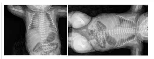
Figure 1 – Thoracoabdominal radiograph performed at birth, showing a radiopaque infradiaphragmatic image on the left, with blurring of the diaphragm, associated with a radiolucent paravertebral image on the right
Case report
Submitted: 24/06/2025
Approved: 04/08/2025
DOI: http://dx.doi.org/10.31365/issn.2595-1769.2026.0374
Infradiaphragmatic Bronchopulmonary Sequestration In A Premature Newborn: Case Report
Sequestro Broncopulmonar Infradiafragmático Em Recém-Nascido Prematuro: Relato De Caso
Secuestro Broncopulmonar Infradiafragmático En Un Recién Nacido Prematuro: Reporte De Un Caso
Mariana Fabrini Gomes1,2
Gabriel Gamba Pioner1,3
Janaina Cruciani Soldateli1,4
1 Universidade Federal de Santa Catarina, Pediatria – Florianópolis-SC, Brazil.
2 ORCID: https://orcid.org/0000-0001-7974-2063
3 ORCID: https://orcid.org/0009-0003-0305-0206
4 ORCID: https://orcid.org/0009-0005-7801-8782
Corresponding author:
Mariana Fabrini Gomes1
E-mail: marifabrinigomes@gmail.com
ABSTRACT
Introduction: Pulmonary sequestration is a rare disease, which can occur intra or extralobar. It is usually asymptomatic, has good prognosis, and should be suspected in prenatal period. Objective: To document the clinical manifestations, diagnosis, treatment and outcomes of a newborn with infradiaphragmatic pulmonary sequestration, whose suspected diagnosis occurred during the intrauterine period. Case description: Male, late preterm, son of a primiparous mother without comorbidities. An ultrasound performed at 18 weeks and 1 day showed a homogeneous echogenic image on the left, suggestive of pulmonary sequestration. He was born by cesarean section, hypotonic and in apnea, and the umbilical cord was immediately clamped. In neonatal intensive care unit, a chest X-ray was performed and showed a hyperechoic image at the left lung base and deviation in diaphragmatic line. An abdominal ultrasound with Doppler was indicated and performed, showing a suggestive infradiaphragmatic pulmonary sequestration image. The patient was monitored by the surgical team during intra and extra-hospital period. At six months old he underwent computed tomography angiography, which confirmed the diagnosis. At eight months old, he underwent surgical resection, which occurred successfully. Discussion: Infradiaphragmatic pulmonary sequestration - patients type - accounts for less than 1.7% of total. It has an unknown etiology, communicates anomalously with tracheobronchial tree, and has its own blood supply. When clinically present, it manifests in the first months of life, and there is no need for immediate intervention. The diagnosis should be suspected prenatally and treatment usually occurs up to nine months of life.
Keywords: Bronchopulmonary Sequestration; Lung Diseases; Infant, Newborn; Diseases; Infant; Premature; Ultrasonography; Prenatal.
RESUMO
Introdução: O sequestro pulmonar é uma doença rara, podendo ocorrer de maneira intra ou extralobar. Costuma ser assintomática, possui bom prognóstico, e deve ser suspeitada no período pré-natal. Objetivo: Documentar as manifestações clínicas, diagnóstico, tratamento e desfechos de um recém-nascido com sequestro pulmonar infradiafragmático, cuja suspeita diagnóstica ocorreu ainda no período intraútero. Descrição do caso: masculino, prematuro tardio, filho de mãe primigesta, sem comorbidades. Na ultrassonografia feita com 18 semanas e 1 dia, visualizou-se imagem ecogênica homogênea à esquerda, sugestiva de sequestro pulmonar. Nasceu de parto cesáreo, hipotônico e em apneia, clampeamento imediato do cordão umbilical. Em unidade de terapia intensiva neonatal, foi realizada radiografia de tórax, que evidenciou imagem radiopaca em base de pulmão esquerdo e desvio na linha diafragmática, indicada e realizada ultrassonografia abdominal com Doppler, mostrando imagem sugestiva de sequestro pulmonar infradiafragmático. O paciente foi acompanhado pela equipe cirúrgica no período intra e extra-hospitalar. Com seis meses de vida, realizou angiotomografia computadorizada, confirmando o diagnóstico. Aos oito meses, realizou a ressecção cirúrgica, a qual ocorreu sem intercorrências. Discussão: Os casos de sequestro pulmonar infradiafragmáticos são responsáveis por menos de 1,7% do total, sendo este o tipo do paciente aqui descrito. Tem etiologia desconhecida, se comunica de maneira anômala com a árvore traqueobrônica e possui suprimento sanguíneo próprio. A clínica, quando presente, se manifesta nos primeiros meses de vida, não havendo necessidade de intervenção imediata. O diagnóstico deve ser suspeitado no pré-natal, e o tratamento costuma ocorrer até os nove meses de vida.
Palavras-chave: Sequestro Broncopulmonar; Pneumopatias; Recém-Nascido; Recém-Nascido Prematuro; Ultrassonografia Pré-Natal.
Resumen
Introducción: El secuestro pulmonar es una enfermedad rara que puede ocurrir intra o extralobar. Generalmente es asintomático, tiene buen pronóstico y debe sospecharse prenatalmente. Objetivo: Documentar las manifestaciones clínicas, el diagnóstico, el tratamiento y la evolución de un recién nacido con secuestro pulmonar infradiafragmático, cuya sospecha diagnóstica ocurrió durante el período intrauterino. Descripción del caso: Niño, prematuro tardío, hijo de madre primigesta, sin comorbilidades. Una ecografía realizada a las 18 semanas y 1 día mostró una imagen ecogénica homogénea a la izquierda, sugestiva de secuestro pulmonar. Nació por cesárea, hipotónico y apneico, con pinzamiento inmediato del cordón umbilical. En la unidad de cuidados intensivos neonatales, se realizó una radiografía de tórax, que mostró una imagen radiopaca en la base del pulmón izquierdo y desviación en la línea diafragmática. Se indicó y realizó una ecografía abdominal con Doppler, que mostró una imagen sugestiva de secuestro pulmonar infradiafragmático. El equipo quirúrgico monitorizó al paciente tanto intra como extrahospitalario. A los seis meses de edad, una angiografía por tomografía computarizada confirmó el diagnóstico. A los ocho meses, se realizó una resección quirúrgica sin complicaciones. Discusión: Los casos de secuestro pulmonar infradiafragmático representan menos del 1,7 % del total, y este es el tipo de paciente descrito aquí. Presenta una etiología desconocida, se comunica de forma anómala con el árbol traqueobronquial y posee su propio aporte sanguíneo. Cuando se presenta, los síntomas clínicos se manifiestan en los primeros meses de vida y no es necesaria una intervención inmediata. El diagnóstico debe sospecharse prenatalmente y el tratamiento suele administrarse hasta los nueve meses de edad.
Palabras clave: Secuestro broncopulmonar; Enfermedades pulmonares; Recién nacido; Enfermedades; Prematuro; Ultrasonografía prenatal.
Introduction
Pulmonary sequestration is a rare disease,1 described in the literature since 1777, initially as an anomalous pulmonary drainage. Only in 1946 the term “pulmonary sequestration” came to be understood as a clinical entity2,3 and it has been used to the present day. The incidence in newborns (NB) is less than 2%,5,8 totaling up to 6.4% of pulmonary malformations, being the second most common condition in this group of diseases.6 These values may be underestimated, since some adults present complications of the disease without prior diagnosis, or remain asymptomatic throughout their lives.6 Extralobar cases account for approximately 90% of diagnoses made in the prenatal period,11 and rarely present symptomatically during the perinatal period.8 It is a non-functional pulmonary parenchymal mass in which communication with the tracheobronchial tree is anomalous. Its blood supply also does not follow standard anatomy, generally originating from a branch of the descending aorta.2
The clinical presentation of pulmonary sequestration, when present, manifests in the first months of life and occurs with dyspnea, persistent cough, hemoptysis, chest pain, and recurrent respiratory infections.2,9 Its prenatal diagnosis should be suspected in cases of polyhydramnios, hydrops, or a pulmonary mass identified on Doppler ultrasound (USG) between the 18th and 19th week of gestation.3 In the postnatal period, thoracoabdominal USG, chest tomography, magnetic resonance imaging, or angiography elucidate the diagnosis.
The indicated treatment is surgical resection of the pulmonary sequestration or the affected pulmonary segment, which can be performed via thoracotomy or video-assisted thoracoscopic surgery (VATS), especially in symptomatic patients.2,3 It is a disease with a good prognosis and an important differential diagnosis of recurrent pneumonia and persistent symptoms such as cough and dyspnea.
Case description
Preterm male newborn with a gestational age (GA) of 35 weeks and 1 day and a birth weight of 3,340 g. Son of a primigravida mother, 37 years old, healthy, who had eight prenatal visits. Maternal serologies were negative with non-reactive rapid tests for syphilis, HIV and hepatitis B. In the morphological ultrasound at GA of 18 weeks and 1 day, a homogeneous echogenic image measuring 37x23x35mm was visualized in the thoracoabdominal transition region on the left, suggestive of pulmonary sequestration.
The newborn was born by cesarean section due to premature rupture of membranes and breech presentation. He was born hypotonic and apneic, the cord was immediately clamped, and he was taken to a heated crib. After the initial steps of neonatal resuscitation and one cycle of positive pressure ventilation, he showed improvement in tone and spontaneous breathing. Apgar 7/8. The patient developed respiratory distress (grunting) and decreased oxygen saturation, requiring CPAP for approximately 3 minutes, followed by improvement. He was transferred to the Neonatal Intensive Care Unit (NICU), totaling 7 days of hospitalization. He did not require ventilatory support or oxygen during his stay.
In the NICU, a chest X-ray was performed (Figure 1), revealing a radiopaque image at the lower base of the left lung, with deviation in the diaphragmatic line. Due to the radiological alteration and previous obstetric ultrasound, an abdominal ultrasound with Doppler was performed, which identified a homogeneous echogenic mass with well-defined contours located in the infradiaphragmatic region, paramedian to the left, in contact with the adrenal gland on the same side and displacing the aorta to the contralateral side, measuring approximately 5.0 x 3.4 x 2.5 cm, well vascularized, with irrigation from a branch of the aorta, suggestive of infradiaphragmatic pulmonary sequestration.
Figure 1 – Thoracoabdominal radiograph performed at birth, showing a radiopaque infradiaphragmatic image on the left, with blurring of the diaphragm, associated with a radiolucent paravertebral image on the right
The case was discussed with the surgical team on the patient’s first day of life, and since the respiratory distress presented by the newborn was not related to the surgical disease, there was no indication for immediate surgical intervention. He was discharged from the neonatal unit with a referral to the pediatric surgery team of a regional referral hospital for surgical planning according to clinical conditions, and high-risk outpatient follow-up, in addition to referral to an orthopedist due to congenital clubfoot.
At 6 months of age, he underwent computed tomography angiography (CT angiography) of the chest (Figure 2), which showed signs consistent with pulmonary sequestration along the posterior basal segment of the left lower pulmonary lobe, with infradiaphragmatic extension, fed by an artery originating from the abdominal aorta (Figure 2), and drainage with an extremity between the splenic and portal veins.
At 8 months of age, he underwent resection of the pulmonary sequestration by video-assisted thoracoscopy. During the procedure, bulging of the abdominal diaphragm was observed, leading to the decision to convert the surgery to an open approach, which was performed via laparotomy. A large retroperitoneal vascularized lesion was identified and completely removed. The procedure was uneventful, and the patient showed good neuropsychomotor development and good weight gain.
Figure 2 – Computed tomography angiography - pulmonary sequestration evidenced and its vascularization in the mediastinal window
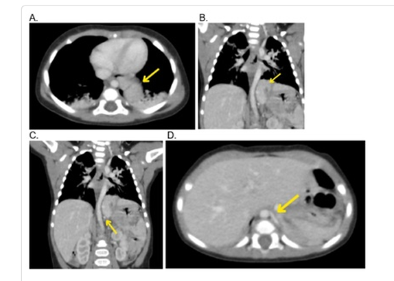
A. pulmonary (intralobar) portion B. infradiaphragmatic (extralobar) portion C and D. vascularization originating from the abdominal aorta towards the pulmonary sequestration.
Discussion
Pulmonary sequestration is a rare disease. In newborns, its incidence is less than 2%,5,8 with extralobar cases accounting for about a quarter of diagnoses. Among these, infradiaphragmatic pulmonary sequestrations, such as the one described above, have an even lower incidence, ranging from 0.5 to 1.7%.2,3,11
Its etiopathogenesis is unknown, but the main theories state that it originates in the primitive gut, from the formation of an extranumerary accessory lung that migrates caudally, along with the esophagus, as in extralobar cases.2,3,10 It is a non-functional pulmonary parenchymal mass whose communication with the tracheobronchial tree is anomalous. Its blood supply does not follow standard anatomy and is usually derived from a branch of the descending aorta.2 In the case described above, as visualized in the chest CT angiography, the mass presented rich vascularization, requiring meticulous anatomical study through imaging to define the best surgical approach.
The disease is classified as intralobar and extralobar. This division is made based on the presence or absence of a layer of the visceral pleura proper surrounding the anomalous cyst.4 When present, it corresponds to the intralobar form and, when absent, to the extralobar form.2 The extralobar form can also present in an infradiaphragmatic manner, an even rarer form.
The intralobar form corresponds to 75 to 85% of cases and is rarely diagnosed in fetal life. It is more common in the left lower pulmonary lobe. The extralobar form accounts for 15 to 25% of cases2,3,11 and is frequently associated with other congenital malformations.4
Extralobar cases, which account for about 90% of diagnoses made in the prenatal period,11 rarely present with symptoms during the perinatal period.8 This fact justifies the lack of surgical intervention on the first day of life, when the patient presented with dyspnea. Pulmonary sequestration predominates in males, with a ratio of 4 cases to 1 in females.10 It is frequently associated with other malformations, especially diaphragmatic hernia, as well as cystic adenomatoid malformation (CAM), bronchogenic cyst, cardiovascular anomalies, pulmonary hypoplasia, fetal hydrops, and pectus excavatum.2,10
The clinical presentation of pulmonary sequestration, when present, manifests in the first months of life and occurs with dyspnea, persistent cough, hemoptysis, chest pain, and recurrent respiratory infections.2,9 The patient reported here did not present compatible clinical symptoms, having good neuropsychomotor and weight-for-height development throughout the months, and an absence of recurrent infectious episodes. He only presented dyspnea at birth, which was more associated with prematurity than with pulmonary sequestration.
Prenatal diagnosis should be suspected in cases of polyhydramnios, hydrops, or a pulmonary mass identified on Doppler ultrasound (USG) between the 18th and 19th week of gestation,3 where a homogeneous or heterogeneous, hyperechoic, circumscribed solid mass of varying location is visualized, in addition to assessing gestational age and blood supply.2,5,6 In some cases, consolidation, multiple cystic masses with air-fluid levels, cavitary lesions, or a single lesion may also be seen.1 Rarely, patients remain beyond the first decade of life without diagnosis and adequate treatment.9,1 In the case described, the mass was evidenced early and the suspicion correctly raised, but the Doppler examination was not performed prenatally.
In the neonatal period, thoracoabdominal ultrasound is usually the first examination requested, as is done in newborns, identifying a hyperechoic image, which may have a thin hyperechoic halo.5 In older children, a simple chest X-ray may indicate the diagnosis, with the presence of a homogeneous mass.2 However, confirmation is made by chest computed tomography, magnetic resonance imaging, or angiography, the latter being the gold standard.2 The need for biopsy is controversial, since the risk of bleeding is high.3
The recommended treatment is surgical resection of the pulmonary sequestration or the affected pulmonary segment, which can be performed via thoracotomy or video-assisted thoracoscopic surgery (VATS), especially in symptomatic patients.2,3 Conservative approaches, in addition to having no therapeutic effect, increase the chance of complications and risks associated with pulmonary sequestration.7 Lobectomy is the treatment of choice when pulmonary sequestration and its drainage are well established.3 Furthermore, there are cases described in the literature of endovascular treatment, through embolization of the anomalous vasculature, leading to a reduction in blood supply to the mass and consequent tissue necrosis.3 This procedure has a lower incidence of complications and lower morbidity, but more practice and studies are still needed.2,3 This therapy allows for the correction of the pulmonary sequestration itself, as well as other possible concomitant malformations, and reduces the risk of malignant evolution of the mass,5 enabling a better prognosis for the patient.
In the case described, the time between diagnosis and surgical treatment was 8 months, close to the time limit predicted in the literature - between 3 and 9 months12 -, without damage to the patient’s development. As recommended in the literature,2,3 the initial therapeutic option was VATS, a less invasive technique; however, due to the extent and vascularization of the non-functioning lung mass, it was decided to change the surgical method, and laparotomy was performed. The procedure was successfully performed and the patient progressed with complete recovery, without impact on his neuropsychomotor and weight development, possible complications in patients with pulmonary sequestration.13
Pulmonary sequestration has a good prognosis. However, when the patient presents with fetal hydrops or pulmonary hypoplasia, or even extralobar infradiaphragmatic forms, the prognosis may be impaired.3 Some complications are associated with this disease, such as developmental delay, recurrent pulmonary infections, mainly by Pseudomonas aeruginosa, and development of malignant tumors, such as adenocarcinoma.3,13
References
1. Aryal K, Regmi PR, Adhikari G, Karki R, Dhakal P. Intralobar pulmonary sequestration in 2 extremes of ages: A Case report. Radiol Case Rep. 2023 Sep 14;18(11):4145-48. Doi: 10.1016/j.radcr.2023.08.089. PMID: 37745759; PMCID: PMC10511722 Disponível em: https://www.ncbi.nlm.nih.gov/pmc/articles/PMC10511722/
2. Belczak SQ et al. Sequestro pulmonar e tratamento endovascular: um relato de caso. J Vascular Brasil. 2019; 18: e20180110. Disponível em: https://www.scielo.br/j/jvb/a/8bYk9HzMVPW47RCtBsH4Tvx/
3. Chakraborty RK, Modi P, Sharma S. Pulmonary Sequestration. 2023 Jul 24. In: StatPearls [Internet]. Treasure Island (FL): StatPearls Publishing; 2024 Jan. PMID: 30335347. Disponível em: https://www.ncbi.nlm.nih.gov/books/NBK532314/
4. Costa MR et al. Apresentação atípica de sequestro pulmonar extralobar intra-abdominal detectado no pré-natal: relato de caso. Rev Paulista Pediatria. 2016; 34: 243-46. Disponível em: https://www.scielo.br/j/rpp/a/rZ8RCRjtJFCSbFNHPG9ymry/?format=pdf&lang=pt
5. Pêgo-Fernandes PM. et al. Sequestro pulmonar: uma série de nove casos operados. J Pneumol. 2002; 28: 175-79. Doi: https://doi.org/10.1590/S0102-35862002000400001
6. Gabelloni M, Faggioni L, Accogli S, Aringhieri G, Neri E. Pulmonary sequestration: What the radiologist should know. Clin Imaging. 2021 May;73:61-72. Doi: 10.1016/j.clinimag.2020.11.040. Epub 2020 Dec 3. PMID: 33310586. Disponível em: https://www.sciencedirect.com/science/article/pii/S0899707120304885?via%3Dihub
7. Torres Escobar JR, Santa Cruz Estigarribia TJ. Secuestro pulmonar extralobar infradiafragmático: a proposito de un caso. Rev del Nacional (Itauguá). 2015;7(1):50-2. Disponível em: https://www.researchgate.net/publication/283477515_Secuestro_pulmonar_extralobar_infradiafragmatico_a_proposito_de_un_caso
8. Ram A, Warsha F, Vinisha F, Kumar D, Kumari M. Pulmonary Sequestration Mimicking Symptoms of Ischemic Heart Disease. Cureus. 2023 Jun 9;15(6):e40200. Doi: 10.7759/cureus.40200. PMID: 37435277; PMCID: PMC10330954 Disponível em: https://www.ncbi.nlm.nih.gov/pmc/articles/PMC10330954/
9. Sousa V, Carvalho L. Características morfológicas do sequestro pulmonar. Rev Portuguesa Pneumol. 2003; 9(2):153-60. Disponível em: https://www.journalpulmonology.org/pt-caracteristicas-morfologicas-do-sequestro-pulmonar-articulo-S0873215915306656
10. Correia M et al. Sequestro pulmonar intralobar com malformação cardíaca associada. 2015. Doi: https://ojs.pjp.spp.pt/article/view/4135
11. Liu X, Wu R, Zhu S, Gu L, Tang Z. Imaging and pathological characteristics, treatment, and prognosis of pulmonary sequestration-A retrospective study of 13 cases. Clin Respir J. 2023 Sep;17(9):865-873. Doi: 10.1111/crj.13672. Disponível em: https://www.ncbi.nlm.nih.gov/pmc/articles/PMC10500327/
12. Andrade CF, Ferreira HPC, Fischer GB. Congenital lung malformations. J Bras Pneumologia. 2011;37: 259-71. Doi: https://doi.org/10.1590/S1806-37132011000200017
13. Belchis D, Cowan M, Mortman K, Rezvani B. Adenocarcinoma arising in an extralobar sequestration: a case report and review of the literature. Lung Cancer. 2014 Apr;84(1):92-5. Doi: 10.1016/j.lungcan.2014.01.025. Disponível em: https://www.sciencedirect.com/science/article/pii/S0169500214000610?via%3Dihub
Scientific Editor:
Fernanda Pinto Mariz. ORCID: https://orcid.org/0000-0002-6981-2352
Publisher: Sociedade de Pediatria do Rio de Janeiro – SOPERJ
E-mail: secretaria@soperj.org.br
Financial support:
None.
Availability of research data:
The underlying content of the research text is contained in the article.
Conflict of interests:
None.
Authors’ Contributions
MF Gomes: data collection, writing - preparation of the original manuscript, writing - revision and editing.
GG Pioner: writing - review and editing, supervision.
JC Soldateli: methodology, writing - preparation of the original manuscript, writing - revision and editing, supervision.
Rev Pediatria SOPERJ 2026;26(1): e20260374