DOI: http://dx.doi.org/10.31365/issn.2595-1769.2026.0351
Tetrasomy 9p: case report of an infant with favorable outcome
Tetrassomia 9p: relato de caso de lactente com evolução favorável
Tetrasomía 9P: Informe de caso de un lactante con evolución favorable
Anna Jamylle Dias Borges Leal 1
Carlos Henrique Paiva Grangeiro 2
1 Hospital Universitário Walter Cantídio (CH-UFC), Pediatria - Fortaleza - Ceará-Brazil. ORCID: https://orcid.org/0009-0007-3113-4895
2 Hospital Universitário Walter Cantídio (CH-UFC), Genética Médica - Fortaleza - Ceará-Brazil. ORCID: https://orcid.org/0000-0002-4150-7612
Corresponding author:
Anna Jamylle Dias Borges Leal
E-mail: annajamylledias@gmail.com
Submitted: 13/10/2024
Approved: 30/06/2025
Abstract
Introduction: Tetrasomy 9p occurs due to the presence of an isochromosome derived from the short arm of chromosome 9, evidencing a wide range of phenotypic manifestations ranging from craniofacial malformations to incompatibility with life. Objective: To describe an infant with tetrasomy 9p without mosaicism and a favorable clinical evolution despite complications during hospitalization, to add this disease to the list of differential syndromic diagnoses to be considered by the pediatrician. Case description: This is a 7-month-old infant, born to a 32-year-old non-consanguineous couple. During prenatal care, abnormalities were identified on morphological ultrasound, which at birth presented as brachycephaly, ocular hypertelorism, high nasal root (Greek warrior helmet), bilateral preauricular skin tag, blepharophimosis, bilateral cleft lip and palate, atypical genitalia with bilateral cryptorchidism and hypoplasia of the scrotum (Prader 4), ostium secundum-type atrial septal defect (ASD) with hemodynamic repercussions, as well as supratentorial ventricular asymmetry on neuroimaging. The karyotype showed [47, XY, +i(9)(p10)]. Despite multiple complications during prolonged hospitalization, the patient was discharged after 5 months. Discussion: Although it is a rare chromosomal disorder, tetrasomy 9p should be considered by the pediatrician as a differential diagnosis, as its spectrum of presentation is polymorphic and similar to that of other phenotypes of chromosomal anomalies. Despite the unfavorable prognosis, it is possible to de-hospitalize these patients, and the attending physician should be prepared to continue the outpatient follow-up in conjunction with the multidisciplinary team.
Keywords: Tetrasomy; Congenital Abnormalities; Craniofacial Abnormalities; Mosaicism.
RESUMO
Introdução: A tetrassomia 9p ocorre através da presença de um isocromossomo derivado do braço curto do cromossomo 9, evidenciando uma ampla gama de manifestações fenotípicas que vão desde malformações craniofaciais até incompatibilidade com a vida. Objetivo: Descrever um lactente com tetrassomia 9p sem mosaicismo e evolução clínica favorável, apesar de intercorrências durante internação, com o intuito de acrescentar tal doença à lista de diagnósticos sindrômicos diferenciais a ser considerado pelo pediatra. Descrição do caso: Trata-se de um lactente de 7 meses, filho de casal de 32 anos não consanguíneo. Durante o pré-natal, foram identificadas anormalidades em ultrassonografia morfológica que ao nascimento se apresentaram como braquicefalia, hipertelorismo ocular, raiz nasal alta (capacete grego), apêndice auricular bilateral, blefarofimose, fenda labiopalatina bilateral, genitália atípica com criptorquidia bilateral e hipoplasia de bolsa escrotal (Prader 4), comunicação interatrial tipo ostium secundum com repercussão hemodinâmica, além de assimetria ventricular supratentorial em neuroimagem. Realizou cariótipo que demonstrou [47, XY, +i(9)(p10)]. Apesar de múltiplas intercorrências durante internação prolongada, foi possível alta hospitalar após cerca de 5 meses de internação. Discussão: Mesmo sendo uma cromossomopatia rara, a tetrassomia 9p deve ser considerada pelo pediatra como diagnóstico diferencial, já que seu espectro de apresentação é polimórfico e semelhante a outros fenótipos de anomalias cromossômicas. Apesar do prognóstico desfavorável, é possível a desospitalização desses pacientes, devendo o médico assistente estar preparado para dar continuidade ao seguimento ambulatorial em conjunto com equipe multidisciplinar.
Palavras-Chave: Tetrassomia; Anormalidades Congênitas; Anormalidades Craniofaciais; Mosaicismo.
Resumen
Introducción: La tetrasomía 9p se presenta por la presencia de un isocromosoma derivado del brazo corto del cromosoma 9, presentando una amplia gama de manifestaciones fenotípicas que van desde malformaciones craneofaciales hasta incompatibilidad con la vida. Objetivo: Describir el caso de un lactante con tetrasomía 9p sin mosaicismo y con una evolución clínica favorable, a pesar de las complicaciones durante la hospitalización, con el fin de incluir esta enfermedad en la lista de diagnósticos sindrómicos diferenciales que debe considerar el pediatra. Descripción del caso: Se trata de un lactante de 7 meses, hijo de una pareja no consanguínea, ambos de 32 años. Durante el control prenatal, se identificaron anormalidades en la ecografía morfológica, que al nacer se presentaron como braquicefalia, hipertelorismo ocular, raíz nasal alta (casco griego), apéndice auricular bilateral, blefarofimosis, labio y paladar hendido bilateral, genitales atípicos con criptorquidia bilateral e hipoplasia escrotal (Prader 4), comunicación interauricular tipo ostium secundum con repercusión hemodinámica y asimetría ventricular supratentorial en la neuroimagen. El cariotipo mostró [47, XY, +i(9)(p10)]. A pesar de las múltiples complicaciones durante la hospitalización prolongada, el alta hospitalaria fue posible después de aproximadamente 5 meses. Discusión: Si bien es una anomalía cromosómica rara, la tetrasomía 9p debe ser considerada por el pediatra como un diagnóstico diferencial, ya que su espectro de presentación es polimórfico y similar a otros fenotipos de anomalías cromosómicas. A pesar del pronóstico desfavorable, es posible dar de alta del hospital a estos pacientes y el médico tratante debe estar preparado para continuar el seguimiento ambulatorio en conjunto con un equipo multidisciplinario.
Palabras Clave: Tetrasomía; Anomalías congénitas; Anomalías craneofaciales; Mosaicismo.
Tetrasomy 9p occurs through the presence of an isochromosome derived from the short arm of chromosome 9, and is associated with a wide range of phenotypic manifestations, from craniofacial and cardiac malformations to life incompatibility. It was initially reported in 1973 and, being a rare chromosomal anomaly, most cases are diagnosed only after birth, with epidemiology still scarce in the literature.1,2,3
Clinical manifestations are varied and their severity may be related to the presence or absence of mosaicism. Within the spectrum of abnormalities, there are global delay in neuropsychomotor development, intellectual disability, cardiac malformations, genitourinary tract anomalies, intrauterine growth restriction (IUGR), as well as skeletal and ophthalmological alterations. Approximately 20% of cases may present with microcephaly, as well as facial dysmorphisms, external ear abnormalities, ocular hypertelorism, micrognathia, bulbous nasal bridge, and cleft lip and palate.4
We describe the case of a male infant who was diagnosed with tetrasomy 9p without mosaicism in the postnatal period, with multiple alterations identified in morphological ultrasound. He evolved with clinical stability and was discharged from the hospital after about 5 months of hospitalization, despite multiple malformations and complications during the period he was hospitalized in a reference maternity hospital.
A 7-month-old infant, the second child of a 32-year-old mother and the fourth child of a father of the same age, not consanguineous and with no family history of known genetic syndromes. The mother denied using illicit substances during pregnancy, medications, smoking, or alcoholism. Prenatal care was adequately performed, with routine laboratory tests showing no abnormalities. First-trimester ultrasound was within normal limits, showing the presence of the nasal bone, ductus venosus with a positive A wave, and nuchal translucency measuring 1.6 mm. Upon performing a morphological ultrasound, a strawberry-shaped skull, bilateral cleft lip and palate, ocular hypertelorism, enlarged cardiac area associated with pericardial effusion, bilateral grade I pyelocaliceal dilation, and IUGR were demonstrated.
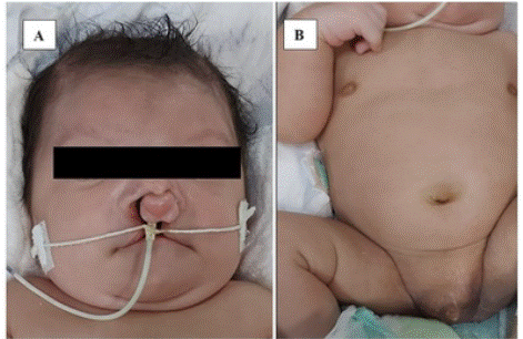
The patient was born at 38 weeks and 1 day of gestational age, via elective cesarean section due to fetal malformation, IUGR, and breech presentation. The infant required neonatal resuscitation in the delivery room and responded well after 2 cycles of positive pressure ventilation (PPV), presenting Apgar scores of 5 and 8. Initial assessment showed a weight of 1,840g (z-score -2.93) and a height of 41cm (z-score -3.62), considered small for gestational age (SGA), in addition to a head circumference (HC) of 30.5cm (z-score -2.47). The infant was described as having a brachycephalic skull and dysmorphic facies, exhibiting ocular hypertelorism, high nasal root (Greek helmet), bilateral auricular appendages, blepharophimosis, bilateral cleft lip and palate, and reduction of fifth fingers, as well as atypical genitalia with bilateral cryptorchidism and scrotal hypoplasia, thus classified as sex undetermined (Figure 1). The infant developed respiratory distress and sucking difficulties, and was transferred to the neonatal ICU, where he remained for 24 hours before being transferred to an intermediate care unit.
He had normal transfontanellar, total abdominal, and urinary tract ultrasounds performed on the day of birth. Non-extended biological neonatal screening (heel prick test) was normal.
At 4 days of age, a transthoracic echocardiogram detected an ostium secundum type atrial septal defect (ASD) measuring 7.9 mm with left-to-right flow, dilation of the right chambers, right ventricular overload and hypertrophy, and pulmonary hypertension. He developed neonatal jaundice, requiring phototherapy for 3 days. He presented with late-onset neonatal sepsis due to Staphylococcus epidermidis, which was adequately treated.
Cytogenetic examination with GTG banding using temporary lymphocyte culture with evaluation of 24 metaphases performed at 11 days of age detected an isochromosome of the short arm of chromosome 9, resulting in a partial tetrasomy of chromosome 9 in all metaphases analyzed [47, XY, +i(9)(p10)].
Ophthalmological evaluation at 13 days of age identified telecanthus and normal fundoscopy. Auditory evaluation with absent bilateral otoacoustic emissions (OAEs), as well as a new test performed at 7 months of age. Brainstem audiometry was not performed. At 22 days of age, moderate laryngotracheomalacia was identified.
As an infant, at about one month of age, a pelvic ultrasound was performed which showed signs suggestive of undescended testicles in the inguinal canals. Computed tomography (CT) scan of the skull showed brachycephaly (IC 93), diffuse enlargement of the fontanelles and cranial sutures, mild supratentorial ventricular asymmetry with prominence on the left, increased retro/infracerebellar cerebrospinal fluid spaces, as well as hypoplasia of the inferior cerebellar vermis and bilateral cleft lip and palate. At 2 months, while still hospitalized, he developed a urinary tract infection caused by Klebsiella pneumoniae, which was treated appropriately. He also presented with edema, pain, and hyperemia in his right leg, with venous Doppler ultrasound identifying deep vein thrombosis affecting segments of the common, deep, and superficial femoral veins on the right, receiving treatment with anticoagulation.
At 2 and a half months, a new ultrasound of the kidneys and urinary tract was performed, which demonstrated ectasia of the renal pelvis and central calyces on the left (UTD P1). A new echocardiogram at 3 months of age identified a patent foramen ovale (PFO) measuring 1.9 mm, with left-to-right flow, without hemodynamic repercussions. Transfontanellar ultrasound performed at 4 months showed asymmetrical dilation of the lateral ventricles, asymmetry of the choroid plexuses, and a small choroid plexus cyst, all located on the left side of the brain.
A gastrostomy was performed around 5 months of age, in addition to a new venous Doppler ultrasound of the right lower limb, which showed no signs of thrombosis. The patient was then discharged from the hospital at 5 months and 9 days of age, clinically stable, for follow-up with a multidisciplinary team and outpatient monitoring. The parents are awaiting outpatient karyotype collection for both.
DISCUSSION
Tetrasomy 9p is a rare chromosomal anomaly first described in 1973, with 72 cases reported up to January 2022. It results from a supernumerary isochromosome formed by two copies of the short arm of chromosome 9, and its severity is related to the presence or absence of mosaicism. The prevalence of supernumerary isochromosomes varies from 0.14 to 0.72 cases per thousand live births, being i(8p), i(9p), i(12p), i(18p), and i(22p) the most described presentations. It is usually diagnosed postnatally, and about 30% of cases present with mosaicism, manifesting more subtle phenotypic alterations and a better prognosis; cases with an absence of clinical manifestations have also been observed in the literature. Cases without mosaicism present with a wide variety of manifestations, some with greater morbidity, such as the case of the reported infant.1,3,5,6,7,8,9,10
Among the main phenotypic alterations involved, IUGR, central nervous system (CNS) and neuropsychomotor development anomalies, skeletal and craniofacial, cardiac and genitourinary alterations can be highlighted. Maternal age does not appear to be associated with this chromosomal abnormality.1,11
Regarding IUGR, the patient in question was SGA and low birth weight, characteristics common in approximately 57% of cases. The presence of delayed neuropsychomotor development may be present in up to 73% of cases, showing itself to be the most common clinical manifestation according to the literature.1,11
Neuroimaging examination identified cranial alterations such as brachycephaly (IC 93) and diffuse enlargement of the fontanelles and cranial sutures. The head circumference was below 2 standard deviations. As for CNS anomalies, there was supratentorial ventricular asymmetry with left prominence, increased retro/infracerebellar cerebrospinal fluid spaces, with hypoplasia of the inferior cerebellar vermis. Ventriculomegaly and Dandy-Walker malformation are the most common neurological manifestations, observed in up to 78% of patients. Microcephaly may be present in about 20% of cases, an alteration present in the reported patient.1,4,11,12
There were significant facial dysmorphisms such as bilateral cleft lip and palate identified on physical examination and neuroimaging, in addition to manifestations observed such as ocular hypertelorism, high nasal root (Greek helmet), bilateral auricular appendage, and blepharophimosis. According to the literature, ocular hypertelorism may be present in 56% of cases and cleft lip and palate in up to 33%.1,11,12
As for congenital heart disease, at birth an ostium secundum type ASD was identified, measuring 7.9 mm with repercussions on cardiac function; however, an echocardiogram performed at 3 months of age showed only a PFO of 1.9 mm without hemodynamic repercussions. Congenital heart malformations are present in up to 29% to 40% of patients.1,11
He presented with atypical genitalia, with bilateral cryptorchidism and scrotal hypoplasia, with the male sex confirmed only after karyotyping. Ultrasound of the urinary tract at birth was normal; however, a new examination performed at 2 and a half months of age demonstrated ectasia of the renal pelvis and central calyces on the left (UTD P1), with urogenital and renal anomalies, such as the presence of renal microcysts, hypospadias and ambiguous genitalia, identified in up to 29 to 45% of cases of tetrasomy 9p already described.11,13,14
The infant presented with telecanthus on ophthalmological evaluation, with the remainder of the examination being normal. He also presented with absent otoacoustic emissions in both the first evaluation and a new test performed at 7 months of age. Auditory and external ear alterations can occur in up to 69% of cases and ocular alterations in up to 43%.1,9
Laryngotracheomalacia was observed, without the need for interventions, a manifestation not seen in the phenotype of the aforementioned chromosomal abnormality. Deep vein thrombosis in a patient was associated with central venous catheter puncture, and no cases of thrombophilia associated with this tetrasomy have been described.15,16
During the 5 months of hospitalization, the infant presented infectious complications that may be associated with the prolonged period in a hospital environment, but screening tests for immunodeficiencies were not performed at first and were scheduled for post-discharge follow-up appointments. Clinical manifestations related to immune system dysregulation, including myositis and systemic lupus erythematosus-like symptoms, have been described in patients with tetrasomy 9p with mosaicism. Increased expression of genes stimulated by type I interferon has been described in these patients, which may justify such manifestations of dysregulation.16
Despite the unfavorable prognosis and rare etiology, tetrasomy 9p without mosaicism should be included in the pediatrician’s list of differential diagnoses regarding chromosomal anomalies, as it shares phenotypes with other syndromes of clinical interest.
Therefore, in addition to a dysmorphological physical examination performed by a qualified professional at the referral maternity hospital, access to complementary genetic tests and genetic counseling is necessary – such as, in the case mentioned, karyotyping. In this way, it is possible to establish early diagnoses and interventions, preparing both the family that will receive this child and the multidisciplinary team that will accompany them throughout their life in a timely manner.
The progressive expansion of access to referral health services and the training of teams allow for greater survival and chances of hospital discharge for these patients, requiring follow-up with a prepared multidisciplinary team from the moment of diagnosis until beyond hospital discharge.
This case highlights the importance of considering tetrasomy 9p as a differential diagnosis and illustrates that, despite multiple malformations, complications, and prolonged hospital stay, hospital discharge is possible for these patients. However, outpatient clinical follow-up with competent professionals is essential, enabling patient care and support for the family.
REFERENCES
1. Süleyman M, Oğuz S, Kaykı G, Çelik HT, Şimsek-Kiper PÖ, Utine GE, et al. A very rare case of a newborn with tetrasomy 9p and literature review. Turk J Pediatr. 2022;64(1):171-8. https://doi.org/10.24953/turkjped.2021.685
2. Yu J, Chen N, Chen M, Shen M, Qian Y, Dong M. Case Report: Prenatal diagnosis of fetal tetrasomy 9p initially identified by non-invasive prenatal testing. Front Genet. 2022 Oct 31;13:1020525. https://doi.org/10.3389/fgene.2022.1020525
3. Ghymers D, Hermann B, Distèche C, Frederic J. Tétrasomie partielle du chromosome 9, à l'état de mosaïque, chez un enfant porteur de malformations multiples. Humangenetik. 1973 Dec 10;20(3):273-82. https://doi.org/10.1007/BF00385740
4. Bellil H, Herve B, Herzog E, Ayoubi JM, Vialard F, Poulain M. A high level of tetrasomy 9p mosaicism but no clinical manifestations other than moderate oligozoospermia with chromosomally balanced sperm: a case report. J Assist Reprod Genet. 2020 Mar;37(3):573-577. https://doi.org/10.1007/s10815-020-01690-0
5. Chen CP, Wang LK, Chern SR, Wu PS, Chen YT, Kuo YL, et al. Mosaic tetrasomy 9p at amniocentesis: prenatal diagnosis, molecular cytogenetic characterization, and literature review. Taiwan J Obstet Gynecol. 2014 Mar;53(1):79-85. https://doi.org/10.1016/j.tjog.2013.12.002
6. El Khattabi L, Jaillard S, Andrieux J, Pasquier L, Perrin L, Capri Y, et al. Clinical and molecular delineation of Tetrasomy 9p syndrome: report of 12 new cases and literature review. Am J Med Genet A. 2015 Jun;167(6):1252-61. https://doi.org/10.1002/ajmg.a.36932
7. Röthlisberger B, Chrzanowska K, Balmer D, Riegel M, Schinzel A. A supernumerary marker chromosome originating from two different regions of chromosome 18. J Med Genet. 2000 Feb;37(2):121-4. https://doi.org/10.1136/jmg.37.2.121
8. Papoulidis I, Kontodiou M, Tzimina M, Saitis I, Hamid AB, Klein E, et al. Tetrasomy 9p mosaicism associated with a normal phenotype in two cases. Cytogenet Genome Res. 2012;136(4):237-41. https://doi.org/10.1159/000337520
9. Shu W, Cheng SSW, Xue S, Chan LW, Soong SI, Kan ASY, et al. First Case Report of Maternal Mosaic Tetrasomy 9p Incidentally Detected on Non-Invasive Prenatal Testing. Genes. 2021; 12(3):370. https://doi.org/10.3390/genes12030370
10. Liehr T, Al-Rikabi A. Mosaicism: Reason for Normal Phenotypes in Carriers of Small Supernumerary Marker Chromosomes with Known Adverse Outcome. A Systematic Review. Front Genet. 2019 Nov 11;10:1131. https://doi.org/10.3389/fgene.2019.01131
11. Vinkšel M, Volk M, Peterlin B, Lovrecic L. A Systematic Clinical Review of Prenatally Diagnosed Tetrasomy 9p. Balkan J Med Genet. 2019 Aug 28;22(1):11-20. https://doi.org/10.2478/bjmg-2019-0012
12. Moczulska H, Pietrusinski M, Zezawska K, Serafin M, Skoczylas B, Jachymski T, et al. Cases of tetrasomy 9p and trisomy 9p in prenatal diagnosis-Analysis of noninvasive and invasive test results. Front Genet 2022 Sep 26;13:994455. https://doi.org/10.3389/fgene.2022.994455
13. Pinto IP, Minasi LB, Steckelberg R, da Silva CC, da Cruz AD. Mosaic Tetrasomy of 9p24.3q21.11 postnatally identified in an infant born with multiple congenital malformations: a case report. BMC Pediatr. 2018 Sep 7;18(1):298. https://doi.org/10.1186/s12887-018-1275-8
14. Wang H, Xie LS, Wang Y, Mei J. Prenatal diagnosis of mosaic tetrasomy 9p in a fetus with isolated persistent left superior vena cava. Taiwan J Obstet Gynecol. 2015 Apr;54(2):204-5. https://doi.org/10.1016/j.tjog.2014.12.005
15. Ions R, Narayanan M, Browning M, Gaillard EA, Stiefel G, Tang JW. Case presentation: persistent adenovirus B3 infections associated with bronchiolitis obliterans treated with cidofovir in a child with mosaic tetrasomy 9p. BMC Infect Dis. 2018 Oct 22;18(1):529. https://doi.org/10.1186/s12879-018-3441-x
16. Frémond ML, Gitiaux C, Bonnet D, Guiddir T, Crow YJ, de Pontual L, et al. Mosaic Tetrasomy 9p: A Mendelian Condition Associated With Pediatric-Onset Overlap Myositis. Pediatrics. 2015 Aug;136(2):e544-7. https://doi.org/10.1542/peds.2015-0724
Figure 1 - Craniofacial and genital dysmorphia observed in the infant.
(A) Bilateral cleft palate. (B) Atypical genitalia with bilateral cryptorchidism and scrotal hypoplasia.
Associate Editor:
Clarisse Pereira Dias Drumond Fortes
ORCID: https://orcid.org/0000-0002-8253-0501:
Publisher: Sociedade de Pediatria do Rio de Janeiro – SOPERJ
E-mail: secretaria@soperj.org.br
Financial support:
None.
Availability of research data:
The underlying content of the research text is contained in the article.
Conflict of interests:
None.
Authors’ contributions
AJDB Leal: data collection, conceptualization, investigation, methodology, writing - preparation of the original, visualization
CHP Grangeiro: conceptualization, project management, methodology, writing - revision and editing, supervision, visualization.
Rev Pediatria SOPERJ 2026;26(1): e20260351