IntroduCTION
Angelman syndrome (AS; OMIM #105830) was first described in 1965 by Harry Angelman, a British pediatrician who reported three children with severe intellectual disability, absence of language, excessive unmotivated laughter, and ataxic movements, whom he called “puppet children”.1 In 1967, Bower and Jeavons described two children whose clinical characteristics included delayed neuropsychomotor development, severe intellectual disability, hypotonia, ataxia, epilepsy, absence of language, and a characteristic face with a large jaw and open mouth.2 Only in 1982 was the term “puppet children” replaced by Angelman syndrome, as the initial name was considered pejorative for the patient and their family.3
AS is a rare neurodevelopmental disorder of genetic origin that occurs due to the loss of function of the maternal copy of the UBE3A gene (UBE3A – OMIM *601623 – Ubiquitin-Protein Ligase E3A – locus 15q11.2),4 responsible for encoding a protein that functions both as an E3 ligase in the ubiquitin proteasome pathway and as a transcriptional coactivator. The UBE3A gene is subject to genomic imprinting, with specific maternal preferential expression in the brain and, more specifically, in neurons, but does not occur in glial cells.5 AS has an estimated incidence of between 1:12,000 and 1:24,000 births.6 In Brazil, the authors did not find epidemiological data regarding AS.
The etiology of AS is not Mendelian, being explained through four distinct molecular mechanisms, which directly involve genetic counseling and recurrence risk, all involving the loss of expression of the maternally inherited UBE3A gene: (1) Deletion of the 15q11.2–q13 region of the maternal chromosome, occurring in approximately 70% of cases;7 (2) Paternal uniparental disomy of chromosome 15, with the imprinting[a] pattern occurring in both critical regions of the paternally inherited chromosome 15, 2 to 3% of cases and, consequently, the UBE3A gene is not expressed, representing approximately 2% of cases;7 (3) Imprinting errors in the 15q11.2-q13 region of the maternally inherited chromosome 15, promoting the non-expression of the UBE3A gene, occurring in 2 to 3% of cases;7,8 and (4) Pathogenic heterozygous variant of the maternally inherited UBE3A gene, occurring in approximately 10% of cases.7
AS is characterized by clinical manifestations that mainly include severe delay in neuropsychomotor development (NPMD), severe intellectual disability, gait ataxia and/or limb incoordination, absence of language, and happy behavior that includes unmotivated and excessive laughter.1,9,10 In addition, microcephaly, seizures, and sleep disorders are quite common.11,12 Other findings that may also be observed are autism spectrum disorder (ASD), hyperactivity, strabismus, macrostomia, spaced teeth, open mouth, protruding tongue, prognathism, sialorrhea, feeding problems, hypotonia during childhood, scoliosis, constipation, attraction/fascination with water.9 In relation to their relatives, hypopigmentation of the skin, hair, and light eyes may occur in cases where there is a chromosomal deletion, leading to haploinsufficiency of the OCA2 gene (located near the gene UBE3A), the OCA2 gene is responsible for encoding a protein involved in tyrosine metabolism that is related to the development of skin, hair, and iris pigmentation.13,14
The aim of this study is to report on a child diagnosed with AS.
CASE DescriPTION
Proband – male, 4 years old. Referred to the Medical Genetics service at 3 years and 11 months of age by the Family Clinic for global delay in neuropsychomotor development.
He is the only child of a non-consanguineous couple, healthy and with no history of other similar cases, malformations or genetic diseases in the family. Mother was 37 years old at the time of pregnancy. Gesta I, Para I. Pregnancy without complications and eight prenatal visits. Denies exposure to known teratogenic agents. Cesarean delivery at 37 weeks, without perinatal asphyxia (Apgar score15 8/9 at the first and fifth minute, respectively). Weighed 2,380g (below p10), measured 47cm (between p3 and p15), head circumference 34cm (between p15 and p50). Small for gestational age.16 No perinatal complications. Normal neonatal screenings. Discharge from the maternity ward at 72 hours of life.
At 15 months of age, the parents reported to the pediatrician that the infant was not speaking or walking, and he was then referred to neuropediatrics and otolaryngology.
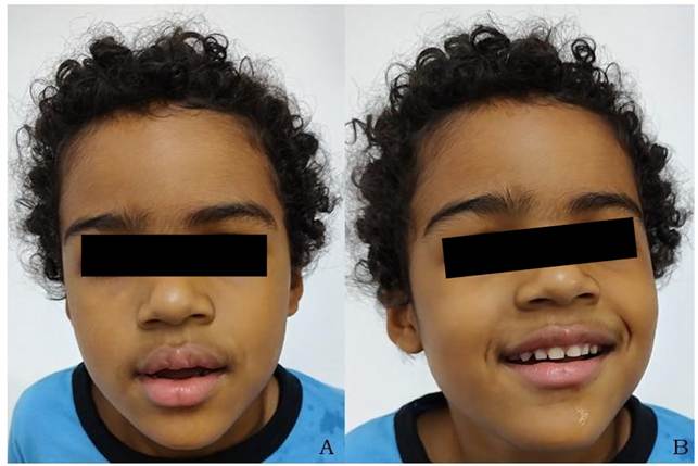
Physical and morphological examination at the first evaluation, in Genetics, showed normal height (50th percentile), normocephaly (between 15th and 50th percentiles). Hypotonia. Black and curly hair, broad forehead, hypertrichosis on the forehead, synophrys, long eyelashes, epicanthus, square nasal base, depressed nasal bridge, short philtrum, macrostomia with open mouth, thick and everted lips, spaced teeth, sialorrhea, prognathism, unmotivated laughter (Figure 1). Ataxic gait on tiptoes, unstable, in a position of flexion of the wrists and elbows. Absence of language, hyperactivity and easily excitable, ASD, sleep-wake cycle alterations, fascination with water and crumpled paper.
Figure 1 - Photos of the proband – broad forehead, hypertrichosis on the forehead, synophrys, short philtrum, thick and everted lips (A). Macrostomia, open mouth, spaced teeth, sialorrhea (B).
Complementary exams: normal magnetic resonance imaging of the skull, cervical spine, thoracic spine and lumbosacral spine. Normal electroencephalogram, brainstem evoked potential, echocardiogram and total abdominal ultrasound. Normal amino acid chromatography. Karyotype 46, XY.20 Molecular research for fragile X syndrome negative. Comparative array genomic hybridization (array-CGH)[b] of peripheral blood revealed a 2,068kb deletion in the 15q11.2-q12 region or arr 15q11.2-q12 (24141964_26210817)x1 encompassing 101 genes, including the UBE3A gene. Methylation analysis using the MLPA (Multiplex Ligation-dependent Probe Amplification)[c] technique of the 15q11.2 region of peripheral blood revealed an altered methylation pattern, detecting only the unmethylated allele, thus confirming the presence of the paternal allele and the absence of the maternal allele in the analyzed chromosomal region.
DiscussION
Chart 1 shows the described phenotype of AS correlated with the phenotype of the proband, who obtained positive findings in more than 50% of the parameters, a child with developmental delay, absence of speech, gait ataxia, typical wrist and elbow flexion position, unmotivated laughter, and facial dysmorphisms characteristic of AS. On the other hand, the proband does not present hypopigmentation of the skin, hair, and iris, which is reported in the literature as observed only in cases of deletion, a molecular mechanism identified in the proband. In this study, however, the deletion did not involve the OCA2 gene (OCA2 – OMIM *611409 – Melanosomal Transmembrane Protein – locus 15q12-q13.1) responsible for encoding a protein that corresponds to the mouse mutant “pink eye dilution” (p). Protein plays a role in regulating the pH of melanosomes.17
Chart 1 – Angelman syndrome phenotype according to the OMIM “Clinical Synopsis”, correlated with the proband’s phenotype.
|
Phenotype |
Proband |
|
Obesity (older children) |
NO |
|
Facial dysmorphia (Head and Neck) Postnatal microcephaly Brachycephaly Occipital plane Occipital sulcus Prognathism Strabismus Ocular hypopigmentation Refractive errors (astigmatism, hyperopia, myopia) Macrostomia Protruding tongue Sialorrhea Spaced teeth |
- - - - + - - NO + - + + |
|
Scoliosis |
NO |
|
Skin hypopigmentation (observed only in cases of deletion) |
- |
|
Neurological (Central Nervous System) Delay at DNPM Severe intellectual disability Absence of speech Ataxia with jerky arm movements March with a broad base Clumsy, unstable Limb tremor Hypotonia Convulsions Hyperreflexia Typical position of flexion of the wrists and elbows Alteration of the sleep-wake cycle Electroencephalographic changes Mild cortical atrophy identified on CT scan or MRI |
+ + + + + + - + - - + + - - |
|
Neurological (Psychiatric and Behavioral) Unmotivated laughter Easily excitable Attraction/fascination with water Attraction/fascination with crumpled items (paper, plastic) |
+ + + + |
Caption: Not observed (NO), Present (+), Absent (-).
AS is a rare neurodevelopmental disorder and usually occurs as a result of a de novo genetic alteration, that is, a sporadic case.18 Thus, the authors emphasize that clinical suspicion of AS should be considered by pediatricians in all children who present with clinical signs including developmental delay, intellectual disability, absence of language, gait ataxia, seizures, and behavioral changes such as unmotivated laughter, ASD, and/or hyperactivity. Considering that the pediatrician is, in most cases, the first medical professional to evaluate children with AS, it is essential that they are attentive to clinical signs suggestive of the syndrome. Given the relevance of early diagnosis of Angelman syndrome for clinical and therapeutic management, it is imperative that the general pediatrician be trained to identify its main signs and symptoms. Diagnostic suspicion allows for timely referral to a medical geneticist, who will perform etiological confirmation through appropriate molecular tests and the early institution of multidisciplinary interventions that will have a positive impact on the functional prognosis and quality of life of the patient and their family.
The reported proband presents the typical AS phenotype, confirmed through molecular tests that initially included the MLPA technique, demonstrating an altered methylation pattern of the 15q11.2 chromosomal region with the detection of only the unmethylated allele. Subsequently, array-CGH was performed, which demonstrated the genetic mechanism involved, identifying a heterozygous deletion in the 15q11.2-q12 region of the maternal chromosome encompassing 101 genes, including the UBE3A gene, which represents most AS cases. Thus, identifying the molecular mechanism responsible for causing AS in the proband was fundamental for conducting genetic counseling for the family and estimating the risk of recurrence, which in this case is less than 1%.19
Therefore, the authors emphasize that for all children suspected of having Angelman syndrome, the first test that should be requested is specific methylation PCR for AS. The deletion of the 15q11.2 chromosomal region can be identified by array-CGH or FISH (Fluorescence In Situ Hybridization).20 On the other hand, karyotyping does not have the resolution capacity to detect chromosomal microdeletions (smaller than 5,000Kb), as was the case with the proband reported in this study, in which a loss of genetic material of 2,068Kb in size was identified.
There is no specific treatment for AS; the therapeutic approach is based on the symptoms presented by the patients. The follow-up should be carried out jointly by doctors from various specialties, and multidisciplinary rehabilitation therapies should ideally begin as early as possible. The proband is monitored by a pediatrician and geneticist, and undergoes therapies including speech therapy, occupational therapy, psychology, psychomotor therapy, and physiotherapy.
ReferENCES
1. Angelman H. 'Puppet children': a report of three cases. Dev. Med. Child Neurol. 7: 681-688, 1965.
2. Bower BD, Jeavons PM. The 'happy puppet' syndrome. Arch. Dis. Child. 42: 298-301, 1967.
3. Williams CA, Frias JL. The Angelman ('happy puppet') syndrome. Am. J. Med. Genet. 11: 453-460, 1982.
4. Matsuura T, Sutcliffe JS, Fang P, Galjaard R-J, Jiang Y, Benton CS, Rommens JM, Beaudet AL. De novo truncating mutations in E6-AP ubiquitin-protein ligase gene (UBE3A) in Angelman syndrome. Nature Genet. 15: 74-77, 1997.
5. Dindot SV, Antalffy BA, Bhattacharjee MB, Beaudet AL. The Angelman syndrome ubiquitin ligase localizes to the synapse and nucleus, and maternal deficiency results in abnormal dendritic spine morphology. Hum. Molec. Genet. 17: 111-118, 2008.
6. Mertz LG, Christensen R, Vogel I, Hertz JM, Nielsen KB, Gronskov K, Ostergaard JR. Angelman syndrome in Denmark. birth incidence, genetic findings, and age at diagnosis. Am J Med Genet A. 2013;161A:2197-203.
7. Kishino T, Lalande M, Wagstaff J. UBE3A/E6-AP mutations cause Angelman syndrome. Nature Genet. 15: 70-73, 1997. Note: Erratum: Nature Genet. 15: 411, 1997.
8. Buiting K, Dittrich B, Gross S, Lich C, Farber C, Buchholz T, Smith E, Reis A, Burger J, Nothen MM, Barth-Witte U, Janssen B, et al. Sporadic imprinting defects in Prader-Willi syndrome and Angelman syndrome: implications for imprint-switch models, genetic counseling, and prenatal diagnosis. Am. J. Hum. Genet. 63: 170-180, 1998.
9. Clayton-Smith J, Laan L. Angelman syndrome: a review of the clinical and genetic aspects. J Med Genet 2003;40:87–95.
10. Van Buggenhout G, Fryns JP. Angelman syndrome (AS, MIM 105830). Eur J Hum Genet 2009;17:1367–1373.
11. Williams CA, Beaudet AL, Clayton-Smith J, et al. Angelman syndrome 2005: updated consensus for diagnostic criteria. Am J Med Genet A 2006; 140:413-418.
12. Bruni O, Ferri R, D’Agostino G, Miano S, Roccella M, Elia M. Sleep disturbances in Angelman syndrome: a questionnaire study. Brain Dev 2004;26:233-240.
13. King RA, Wiesner GL, Townsend D, White JG. Hypopigmentation in Angelman syndrome. Am J Med Genet 1993;46:40-44.
14. Fridman C, Hosomi N, Varela MC, Souza AH, Fukai K, Koiffmann CP. Angelman syndrome associated with oculocutaneous albinism due to an intragenic deletion of the P gene. Am J Med Genet A 2003;119:180-183.
15. Apgar V. A proposal for a new method of evaluation of the newborn infant. Curr Res Anesth Analg. 1953: 260-7.
16. Battaglia F, Lubchenco L. A practical classification of newborn infants by weight and gestational age. J Pedi-atr. 1967: p. 159-63.
17. Yuasa I, Umetsu K, Harihara S, Miyoshi A, Saitou N, Park KS, Dashnyam B, Jin F, Lucotte G, Chattopadhyay PK, Henke L, Henke J. OCA2 481Thr, a hypofunctional allele in pigmentation, is characteristic of northeastern Asian populations. J Hum Genet. 2007;52(8):690-693.
18. Knoll JH, Nicholls RD, Magenis RE, Graham JM Jr, Lalande M, Latt SA. Angelman and Prader-Willi syndromes share a common chromosome 15 deletion but differ in parental origin of the deletion. Am J Med Genet1989;32:285-90.
19. Stalker HJ, Williams CA. Genetic counseling in Angelman syndrome: The challenges of multiple causes. Am. J. Med. Genet. 1998;77:54-59.
20. Pinkel D, Landegent J, Collins C, Fuscoe J, Segraves R, Lucas J, et al. Fluorescence in situ hybridization with human chromosome-specific libraries: detection of trisomy 21 and translocations of chromosome 4. Proc Natl Acad Sci U S A.; 85(23):9138-42; 1988.
Associate Editor:
Clarisse Pereira Dias Drumond Fortes ORCID: https://orcid.org/0000-0002-8253-0501
Publisher: Sociedade de Pediatria do Rio de Janeiro – SOPERJ
E-mail: secretaria@soperj.org.br
Financial support:
None.
Availability of research data:
The underlying content of the research text is contained in the article.
Conflict of interests:
None.
Authors’ Contributions
GC Pinto: conceptualization, drafting - preparation of the original manuscript, drafting - revision and editing.
SR dos Santos: project management, drafting - reviewing and editing, supervision.
